Abstract
Objective—To assess the occurrence of arterial remodelling types and its relation with the severity of luminal stenosis in atherosclerotic coronary arteries. Patients and methods—Twenty one de novo coronary lesions of 20 patients, who were scheduled for percutaneous transluminal coronary angioplasty (PTCA), were investigated with intravascular ultrasound before PTCA. Local arterial remodelling at the lesion site was studied by measuring the cross sectional area circumscribed by the external elastic lamina (EEL) relative to the reference site: (EEL area lesion/reference EEL area) × 100%. Three groups were defined. Group A: relative EEL area of less than 95% (shrinkage), group B: relative EEL area between 95% and 105% (no remodelling), group C: relative increase in EEL area of more than 105% (compensatory enlargement). Results—All three types of remodelling were observed at the lesion site: group A (shrinkage) n = 8, group B (no remodelling) n = 5, group C (compensatory enlargement) n = 8. The mean (SD) relative EEL area at the lesion site in group A and C was 83(9)% and 132(30)%, respectively. In group A, 33% of the luminal area stenosis at the lesion site was caused by shrinkage of the artery. In contrast, group C showed that 87% of the plaque area did not contribute to luminal area stenosis because of compensatory arterial enlargement. Conclusions—These results show that both compensatory enlargement and paradoxical shrinkage occurs in the atherosclerotic coronary artery. Next to plaque accumulation, the type of atherosclerotic remodelling is an important determinant of luminal narrowing. Keywords: coronary arteries; atherosclerosis; remodelling; intravascular ultrasound
Full Text
The Full Text of this article is available as a PDF (132.8 KB).
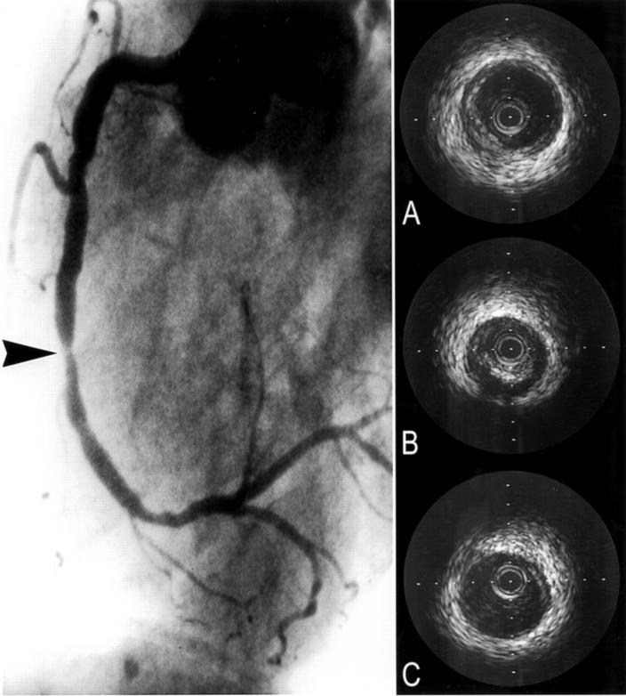
Figure 1 .
Angiogram and intravascular ultrasound images of the right coronary artery in one patient. The arrow indicates the lesion. The intravascular ultrasound images show the proximal reference (A), lesion (B), and distal reference (C) site. At the lesion site arterial shrinkage is present.
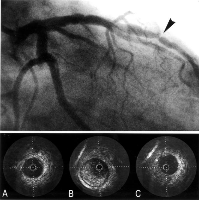
Figure 2 .
Angiogram and intravascular ultrasound images of the left anterior descending coronary artery in another patient. The arrow indicates the lesion. The intravascular ultrasound images show the proximal reference (A), lesion (B), and distal reference (C) site. At the lesion site arterial compensatory enlargement is present.
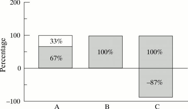
Figure 3 .
This bar graph represents the average percentage plaque contribution (grey bar) and the percentage remodelling contribution (white bar) to the luminal area at the lesion site by intravascular ultrasound in the different three remodelling groups. (A) shrinkage; (B) non-remodelling; (C) compensatory enlargement group. In group A, 67% of the luminal area decrease was contributed by plaque. The remaining 33% luminal area decrease was attributed to paradoxical shrinkage of the lesion. In group B with no remodelling, the stenosis was formed alone by plaque. In group C, plaque contribution was 187% caused by compensatory enlargement of 87% in lumen area.
Selected References
These references are in PubMed. This may not be the complete list of references from this article.
- Eefting F. D., Pasterkamp G., Clarijs R. J., van Leeuwen T. G., Borst C. Remodeling of the atherosclerotic arterial wall: a determinant of luminal narrowing in human coronary arteries. Coron Artery Dis. 1997 Jul;8(7):415–421. doi: 10.1097/00019501-199707000-00003. [DOI] [PubMed] [Google Scholar]
- Glagov S., Weisenberg E., Zarins C. K., Stankunavicius R., Kolettis G. J. Compensatory enlargement of human atherosclerotic coronary arteries. N Engl J Med. 1987 May 28;316(22):1371–1375. doi: 10.1056/NEJM198705283162204. [DOI] [PubMed] [Google Scholar]
- Gussenhoven E. J., Essed C. E., Lancée C. T., Mastik F., Frietman P., van Egmond F. C., Reiber J., Bosch H., van Urk H., Roelandt J. Arterial wall characteristics determined by intravascular ultrasound imaging: an in vitro study. J Am Coll Cardiol. 1989 Oct;14(4):947–952. doi: 10.1016/0735-1097(89)90471-3. [DOI] [PubMed] [Google Scholar]
- Hermiller J. B., Tenaglia A. N., Kisslo K. B., Phillips H. R., Bashore T. M., Stack R. S., Davidson C. J. In vivo validation of compensatory enlargement of atherosclerotic coronary arteries. Am J Cardiol. 1993 Mar 15;71(8):665–668. doi: 10.1016/0002-9149(93)91007-5. [DOI] [PubMed] [Google Scholar]
- Javier S. P., Mintz G. S., Popma J. J., Pichard A. D., Kent K. M., Satler L. F., Leon M. B. Intravascular ultrasound assessment of the magnitude and mechanism of coronary artery and lumen tapering. Am J Cardiol. 1995 Jan 15;75(2):177–180. doi: 10.1016/s0002-9149(00)80072-7. [DOI] [PubMed] [Google Scholar]
- Lamontagne D., Pohl U., Busse R. Mechanical deformation of vessel wall and shear stress determine the basal release of endothelium-derived relaxing factor in the intact rabbit coronary vascular bed. Circ Res. 1992 Jan;70(1):123–130. doi: 10.1161/01.res.70.1.123. [DOI] [PubMed] [Google Scholar]
- Langille B. L., O'Donnell F. Reductions in arterial diameter produced by chronic decreases in blood flow are endothelium-dependent. Science. 1986 Jan 24;231(4736):405–407. doi: 10.1126/science.3941904. [DOI] [PubMed] [Google Scholar]
- Lerman A., Holmes D. R., Jr, Bell M. R., Garratt K. N., Nishimura R. A., Burnett J. C., Jr Endothelin in coronary endothelial dysfunction and early atherosclerosis in humans. Circulation. 1995 Nov 1;92(9):2426–2431. doi: 10.1161/01.cir.92.9.2426. [DOI] [PubMed] [Google Scholar]
- Losordo D. W., Rosenfield K., Kaufman J., Pieczek A., Isner J. M. Focal compensatory enlargement of human arteries in response to progressive atherosclerosis. In vivo documentation using intravascular ultrasound. Circulation. 1994 Jun;89(6):2570–2577. doi: 10.1161/01.cir.89.6.2570. [DOI] [PubMed] [Google Scholar]
- Mintz G. S., Popma J. J., Pichard A. D., Kent K. M., Satler L. F., Wong C., Hong M. K., Kovach J. A., Leon M. B. Arterial remodeling after coronary angioplasty: a serial intravascular ultrasound study. Circulation. 1996 Jul 1;94(1):35–43. doi: 10.1161/01.cir.94.1.35. [DOI] [PubMed] [Google Scholar]
- Nishioka T., Luo H., Eigler N. L., Berglund H., Kim C. J., Siegel R. J. Contribution of inadequate compensatory enlargement to development of human coronary artery stenosis: an in vivo intravascular ultrasound study. J Am Coll Cardiol. 1996 Jun;27(7):1571–1576. doi: 10.1016/0735-1097(96)00071-x. [DOI] [PubMed] [Google Scholar]
- Pasterkamp G., Borst C., Gussenhoven E. J., Mali W. P., Post M. J., The S. H., Reekers J. A., van den Berg F. G. Remodeling of De Novo atherosclerotic lesions in femoral arteries: impact on mechanism of balloon angioplasty. J Am Coll Cardiol. 1995 Aug;26(2):422–428. doi: 10.1016/0735-1097(95)80017-b. [DOI] [PubMed] [Google Scholar]
- Pasterkamp G., Wensing P. J., Post M. J., Hillen B., Mali W. P., Borst C. Paradoxical arterial wall shrinkage may contribute to luminal narrowing of human atherosclerotic femoral arteries. Circulation. 1995 Mar 1;91(5):1444–1449. doi: 10.1161/01.cir.91.5.1444. [DOI] [PubMed] [Google Scholar]
- Post M. J., Borst C., Kuntz R. E. The relative importance of arterial remodeling compared with intimal hyperplasia in lumen renarrowing after balloon angioplasty. A study in the normal rabbit and the hypercholesterolemic Yucatan micropig. Circulation. 1994 Jun;89(6):2816–2821. doi: 10.1161/01.cir.89.6.2816. [DOI] [PubMed] [Google Scholar]
- Post M. J., Borst C., Pasterkamp G., Haudenschild C. C. Arterial remodeling in atherosclerosis and restenosis: a vague concept of a distinct phenomenon. Atherosclerosis. 1995 Dec;118 (Suppl):S115–S123. [PubMed] [Google Scholar]
- Potkin B. N., Bartorelli A. L., Gessert J. M., Neville R. F., Almagor Y., Roberts W. C., Leon M. B. Coronary artery imaging with intravascular high-frequency ultrasound. Circulation. 1990 May;81(5):1575–1585. doi: 10.1161/01.cir.81.5.1575. [DOI] [PubMed] [Google Scholar]
- Wenguang L., Gussenhoven W. J., Zhong Y., The S. H., Di Mario C., Madretsma S., van Egmond F., de Feyter P., Pieterman H., van Urk H. Validation of quantitative analysis of intravascular ultrasound images. Int J Card Imaging. 1991;6(3-4):247–253. doi: 10.1007/BF01797856. [DOI] [PubMed] [Google Scholar]
- Zarins C. K., Weisenberg E., Kolettis G., Stankunavicius R., Glagov S. Differential enlargement of artery segments in response to enlarging atherosclerotic plaques. J Vasc Surg. 1988 Mar;7(3):386–394. [PubMed] [Google Scholar]


