Full Text
The Full Text of this article is available as a PDF (148.7 KB).
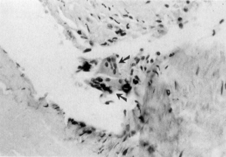
Figure 1 .
Changes after stent implantation showing multinucleate giant cells (arrows). This figure (from von Beusekom1) demonstrates the inflammatory nature of stent implantation, a process that occurs much less after balloon angioplasty. Reprinted from J Am Coll Cardiol 1993;21:45-54 with permission from the American College of Cardiology.
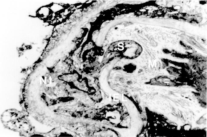
Figure 2 .
Progress of smooth muscle cell (S) from media (M) to new intima (NI) through the damaged internal elastic lamina (IEL) in a balloon injured artery. Once in the new intima the smooth muscle cell proliferates leading to restenosis.
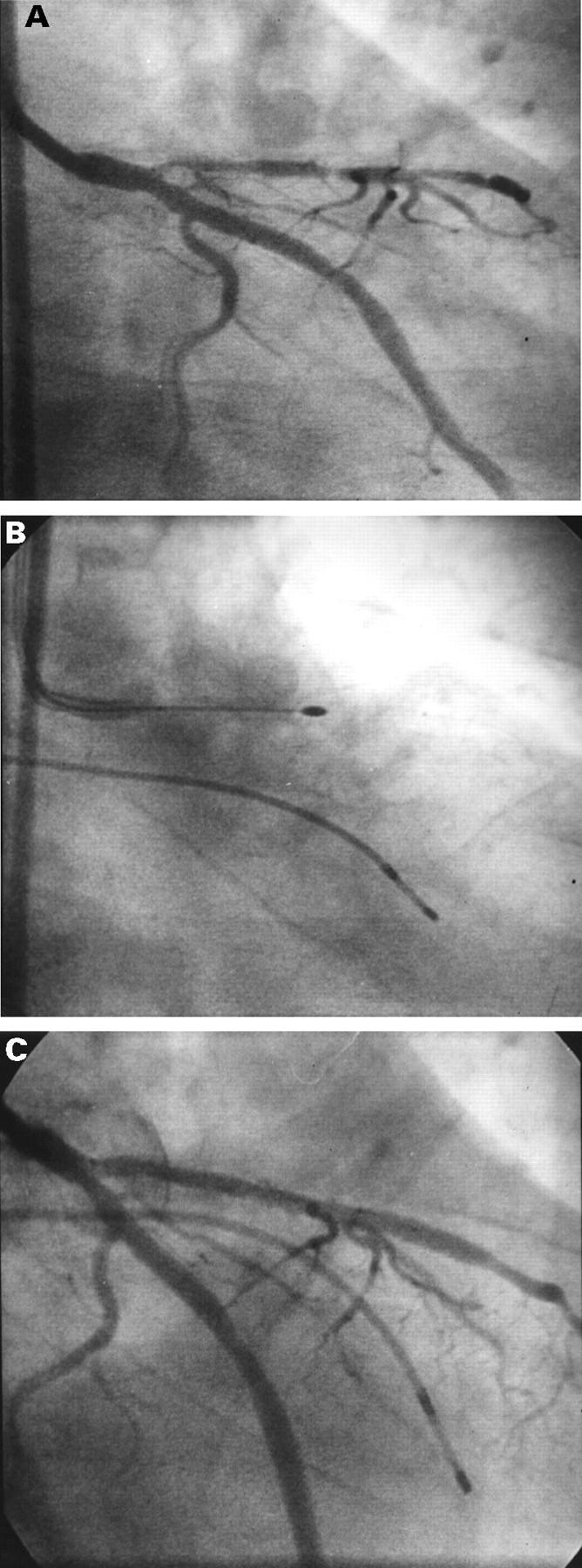
Figure 3 .

(A) Diffuse in-stent restenosis in a Cook GR II stent placed in the left anterior descending artery; (B) rotational atherectomy treatment; (C) final result after rotational atherectomy and ballooning. Regaining the original in-stent luminal diameter can be difficult.
Selected References
These references are in PubMed. This may not be the complete list of references from this article.
- Aggarwal R. K., Ireland D. C., Azrin M. A., Ezekowitz M. D., de Bono D. P., Gershlick A. H. Antithrombotic potential of polymer-coated stents eluting platelet glycoprotein IIb/IIIa receptor antibody. Circulation. 1996 Dec 15;94(12):3311–3317. doi: 10.1161/01.cir.94.12.3311. [DOI] [PubMed] [Google Scholar]
- Baim D. S., Levine M. J., Leon M. B., Levine S., Ellis S. G., Schatz R. A. Management of restenosis within the Palmaz-Schatz coronary stent (the U.S. multicenter experience. The U.S. Palmaz-Schatz Stent Investigators. Am J Cardiol. 1993 Feb 1;71(4):364–366. doi: 10.1016/0002-9149(93)90813-r. [DOI] [PubMed] [Google Scholar]
- Carter A. J., Laird J. R., Bailey L. R., Hoopes T. G., Farb A., Fischell D. R., Fischell R. E., Fischell T. A., Virmani R. Effects of endovascular radiation from a beta-particle-emitting stent in a porcine coronary restenosis model. A dose-response study. Circulation. 1996 Nov 15;94(10):2364–2368. doi: 10.1161/01.cir.94.10.2364. [DOI] [PubMed] [Google Scholar]
- Carter A. J., Laird J. R., Farb A., Kufs W., Wortham D. C., Virmani R. Morphologic characteristics of lesion formation and time course of smooth muscle cell proliferation in a porcine proliferative restenosis model. J Am Coll Cardiol. 1994 Nov 1;24(5):1398–1405. doi: 10.1016/0735-1097(94)90126-0. [DOI] [PubMed] [Google Scholar]
- Dussaillant G. R., Mintz G. S., Pichard A. D., Kent K. M., Satler L. F., Popma J. J., Wong S. C., Leon M. B. Small stent size and intimal hyperplasia contribute to restenosis: a volumetric intravascular ultrasound analysis. J Am Coll Cardiol. 1995 Sep;26(3):720–724. doi: 10.1016/0735-1097(95)00249-4. [DOI] [PubMed] [Google Scholar]
- Fischell T. A., Kharma B. K., Fischell D. R., Loges P. G., Coffey C. W., 2nd, Duggan D. M., Naftilan A. J. Low-dose, beta-particle emission from 'stent' wire results in complete, localized inhibition of smooth muscle cell proliferation. Circulation. 1994 Dec;90(6):2956–2963. doi: 10.1161/01.cir.90.6.2956. [DOI] [PubMed] [Google Scholar]
- Fischell T. A. Polymer coatings for stents. Can we judge a stent by its cover? Circulation. 1996 Oct 1;94(7):1494–1495. doi: 10.1161/01.cir.94.7.1494. [DOI] [PubMed] [Google Scholar]
- King S. B., 3rd Intravascular radiation for restenosis prevention: could it be the holy grail? Heart. 1996 Aug;76(2):99–100. doi: 10.1136/hrt.76.2.99. [DOI] [PMC free article] [PubMed] [Google Scholar]
- Matsuno H., Stassen J. M., Vermylen J., Deckmyn H. Inhibition of integrin function by a cyclic RGD-containing peptide prevents neointima formation. Circulation. 1994 Nov;90(5):2203–2206. doi: 10.1161/01.cir.90.5.2203. [DOI] [PubMed] [Google Scholar]
- Mazur W., Ali M. N., Khan M. M., Dabaghi S. F., DeFelice C. A., Paradis P., Jr, Butler E. B., Wright A. E., Fajardo L. F., French B. A. High dose rate intracoronary radiation for inhibition of neointimal formation in the stented and balloon-injured porcine models of restenosis: angiographic, morphometric, and histopathologic analyses. Int J Radiat Oncol Biol Phys. 1996 Nov 1;36(4):777–788. doi: 10.1016/s0360-3016(96)00298-2. [DOI] [PubMed] [Google Scholar]
- Popowski Y., Verin V., Urban P. Endovascular beta-irradiation after percutaneous transluminal coronary balloon angioplasty. Int J Radiat Oncol Biol Phys. 1996 Nov 1;36(4):841–845. doi: 10.1016/s0360-3016(96)00411-7. [DOI] [PubMed] [Google Scholar]
- Serruys P. W., Emanuelsson H., van der Giessen W., Lunn A. C., Kiemeney F., Macaya C., Rutsch W., Heyndrickx G., Suryapranata H., Legrand V. Heparin-coated Palmaz-Schatz stents in human coronary arteries. Early outcome of the Benestent-II Pilot Study. Circulation. 1996 Feb 1;93(3):412–422. doi: 10.1161/01.cir.93.3.412. [DOI] [PubMed] [Google Scholar]
- Teirstein P. S., Massullo V., Jani S., Popma J. J., Mintz G. S., Russo R. J., Schatz R. A., Guarneri E. M., Steuterman S., Morris N. B. Catheter-based radiotherapy to inhibit restenosis after coronary stenting. N Engl J Med. 1997 Jun 12;336(24):1697–1703. doi: 10.1056/NEJM199706123362402. [DOI] [PubMed] [Google Scholar]
- Thilén U., Aström-Olsson K. Does the risk of infective endarteritis justify routine patent ductus arteriosus closure? Eur Heart J. 1997 Mar;18(3):503–506. doi: 10.1093/oxfordjournals.eurheartj.a015272. [DOI] [PubMed] [Google Scholar]
- Waksman R., Robinson K. A., Crocker I. R., Gravanis M. B., Palmer S. J., Wang C., Cipolla G. D., King S. B., 3rd Intracoronary radiation before stent implantation inhibits neointima formation in stented porcine coronary arteries. Circulation. 1995 Sep 15;92(6):1383–1386. doi: 10.1161/01.cir.92.6.1383. [DOI] [PubMed] [Google Scholar]
- van Beusekom H. M., van der Giessen W. J., van Suylen R., Bos E., Bosman F. T., Serruys P. W. Histology after stenting of human saphenous vein bypass grafts: observations from surgically excised grafts 3 to 320 days after stent implantation. J Am Coll Cardiol. 1993 Jan;21(1):45–54. doi: 10.1016/0735-1097(93)90715-d. [DOI] [PubMed] [Google Scholar]
- van der Giessen W. J., Lincoff A. M., Schwartz R. S., van Beusekom H. M., Serruys P. W., Holmes D. R., Jr, Ellis S. G., Topol E. J. Marked inflammatory sequelae to implantation of biodegradable and nonbiodegradable polymers in porcine coronary arteries. Circulation. 1996 Oct 1;94(7):1690–1697. doi: 10.1161/01.cir.94.7.1690. [DOI] [PubMed] [Google Scholar]


