Abstract
In patients with myocardial bridging, the area within the bridge usually remains free from atherosclerotic disease. The case of a 47 year old man is described who had the rare combination of myocardial bridging with an atherosclerotic plaque within the area of bridging, which was detected with intravascular ultrasound but not with coronary angiography. The clinical history of the patient demonstrates that this is not a benign condition. In symptomatic patients the bridged segment should be screened for the presence of plaque with intracoronary ultrasound. Keywords: myocardial bridging; intravascular ultrasound; atherosclerotic plaque
Full Text
The Full Text of this article is available as a PDF (102.9 KB).
Figure 1 .
Coronary angiogram of the left coronary artery in caudo-cranial projection performed during the first admission. Diastolic (A) and systolic (B) frames demonstrating complete obliteration of the approximately 15 mm long intramural portion of the left anterior descending artery. Arrows indicate the proximal and distal parts of the bridged segment. (C) Occlusion of the left anterior descending artery four weeks later when the patient was readmitted with an acute anterior infarction. The site of the occlusion is in the middle section of the bridge.
Figure 2 .
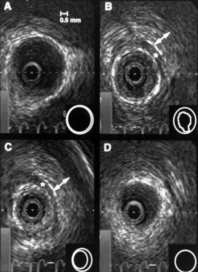
Intracoronary ultrasound images of the patient after PTCA of the left anterior descending artery within the myocardial bridge. A to D represent the course of the left anterior descending artery from proximal to distal. B and C are located within the myocardial bridge. A soft atheromatous plaque was detected that was not apparent on the coronary angiogram. Plaque rupture (B) was probably the result of the PTCA procedure. Asterisks indicate an echolucent band surrounding the artery within the bridge that probably represents adipose tissue around the artery. Vessel lumen and media bounded area are indicated in the insets, and plaque rupture can be seen in panel B at 6 o'clock.


