Abstract
Successful recanalisation of the left anterior descending coronary artery was performed in a 51 year old man who was admitted two weeks after acute anterior myocardial infarction. Fourteen days later, the patient developed Dressler's syndrome with cardiac tamponade, which was immediately punctured. Sternotomy was performed after two weeks because of progressive haemodynamic deterioration, and fibrinous masses were removed from the pericardium. The patient recovered but two weeks later echocardiography showed a perforation of the left ventricular free wall and formation of a pseudoaneurysm. Intensive monitoring showed significant enlargement of the pseudoaneurysm, which was subsequently resected. This case demonstrates that dangerous formation of a pseudoaneurysm can occur not only during the first days of acute myocardial infarction but also after weeks in patients suffering from non-infectious pericarditis caused by Dressler's syndrome. Although the incidence of Dressler's syndrome is declining, patients should be monitored carefully for several weeks, especially by echocardiography. Keywords: Dressler's syndrome; pseudoaneurysm; myocardial infarction
Full Text
The Full Text of this article is available as a PDF (104.6 KB).
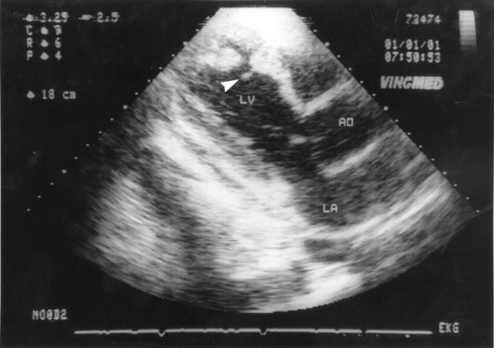
Figure 1 .
Four weeks after the onset of Dressler's syndrome (eight weeks after myocardial infarction) the pseudoaneurysm was primarily detected. Echocardiographic three chamber view shows perforation of the left ventricular wall, extracardial space, and floating parts of myocardial tissue at the margins of the perforation (arrow). LA, left atrium; LV, left ventricle; AO, aorta.
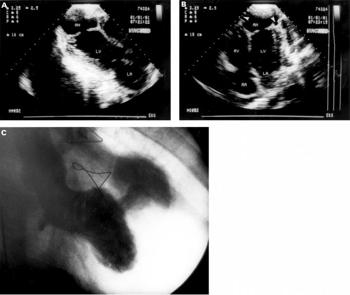
Figure 2 .
Six weeks later (14 weeks after myocardial infarction). (A) Three chamber view shows pericardial effusion, significant enlargement of the perforation, and of the false lumen. (B) Four chamber view shows echo-free edges at the margins of the false lumen (arrows), which was interpreted as progressive detachment of the pasted epicardium and pericardium. (C) Left ventriculography performed preoperatively (right anterior oblique projection) showing the large size of the pseudoaneurysm. LA, left atrium; RA, right atrium; RV, right ventricle; LV, left ventricle; AO, aorta; AN, false lumen of pseudoaneurysm.


