Abstract
A 73 year old man developed a left ventricular pseudoaneurysm following acute myocardial infarction. Coronary angiography showed triple vessel disease with total occlusion of the right coronary artery. On left ventriculography, a serpentine-like pseudoaneurysm was demonstrated that originated from the posterobasal wall of the left ventricle and extended to the right ventricular free wall. He underwent coronary artery bypass surgery with no plication of the pseudoaneurysm. An organised thrombus was also found within the cavity of the pseudoaneurysm. He was doing well approximately eight months after the operation. The prognosis might be determined by the organised thrombus, the serpentine-like structure of pseudoaneurysm, the coronary revascularisation, and the vigorous medical management. Keywords: acute myocardial infarction; pseudoaneurysm; coronary artery bypass surgery
Full Text
The Full Text of this article is available as a PDF (167.7 KB).
Figure 1 .
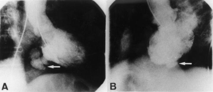
Left ventriculography in the right anterior oblique 30° (A) and left anterior oblique 60° (B) projection. A serpentine-like pseudoaneurysm (arrow) is shown to originate from the left ventricular posterobasal wall and extend to the right ventricular free wall.
Figure 2 .

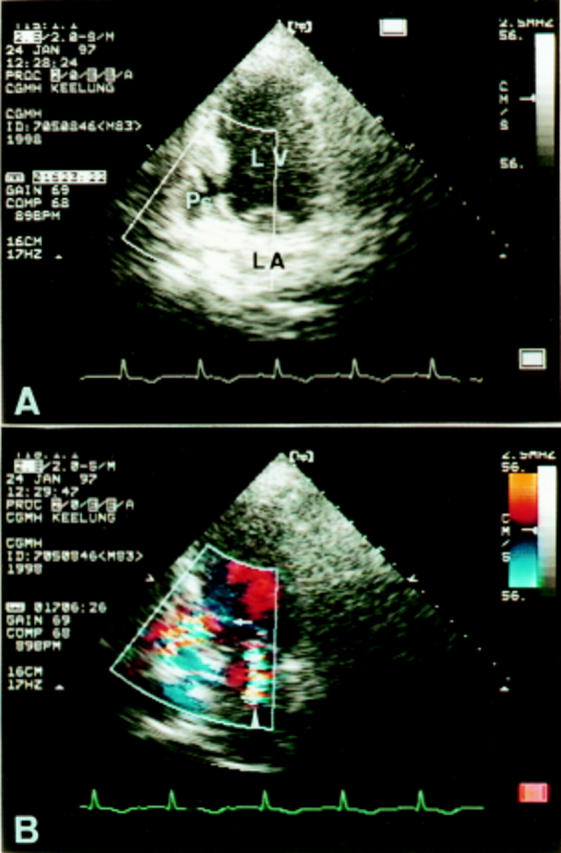
(A) Apical two chamber view of transthoracic cross sectional echocardiography showing an echo free space (pseudoaneurysm) posterior to the left ventricle. (B) Colour Doppler study demonstrating a flow entering the pseudoaneurysm (arrow) and a mitral regurgitant jet (arrowhead). LA, left atrium; LV, left ventricle; Ps, pseudoaneurysm.
Figure 3 .

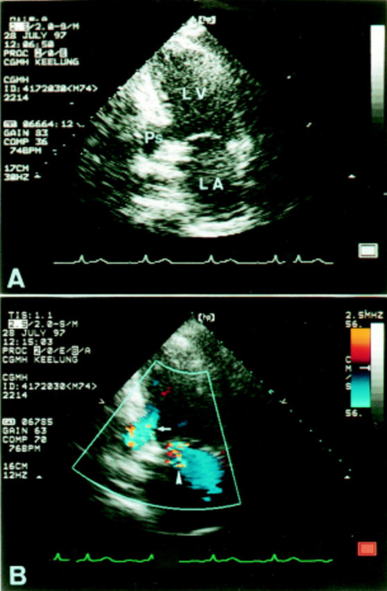
(A) Apical two chamber view of transthoracic cross sectional echocardiography six months after myocardial infarction, showing the pseudoaneurysm posterior to the left ventricle. (B) Colour Doppler study revealing a flow entering the pseudoaneurysm (arrow) and a mitral regurgitant jet (arrowhead). LA, left atrium; LV, left ventricle; Ps, pseudoaneurysm.


