Abstract
Objective—To report initial findings from a selected group of patients with morphological variations of the atrial septal defect who underwent transcatheter closure with a second generation redesigned double umbrella device. Patients—Two patients with abnormal location of the oval fossa and partial deficiency of the septal rim, three patients with multiple defects, and two patients with a multiperforated aneurysm of the interatrial septum (age range, 3.6-25.5 years). Methods—Defects were closed with the double umbrella device (CardioSEAL) consisting of two sets of flexible arms (with central and two mid-arm hinges) covered with sewn Dacron patches. The implantation procedure was monitored by transoesophageal echocardiography. Results—The diameter of the defect measured during transoesophageal echocardiography ranged from 7-18 mm and the balloon stretched diameter ranged from 13-21 mm. The size of the devices varied from 28-33 mm and the ratio of device size to defect size varied from 1.6-2.1. Two devices (23 and 28 mm) were chosen in a patient with two separated defects. No complications or serious arrhythmias were observed during implantation or follow up (median, 1.8 months). Residual shunting was trivial in three patients and mild in one patient (inferiorly located additional defect). Conclusions—To extend the selection critera of an isolated central interatrial defect for transcatheter closure, some modifications of the implantation technique are needed. Using the redesigned double umbrella device, effective closure in patients with multiple or irregularly shaped atrial septal defects was achieved, indicating a broadening of the spectrum of transcatheter closure. Keywords: atrial septal defects; transcatheter closure; congenital heart disorders; double umbrella device; CardioSEAL
Full Text
The Full Text of this article is available as a PDF (134.1 KB).
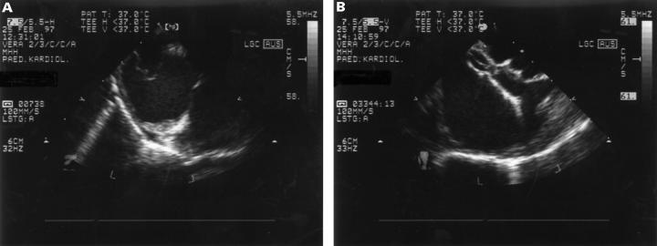
Figure 1 .
Transoesophageal echocardiography (A) before and (B) after occlusion in a patient with a multiperforated aneurysm.
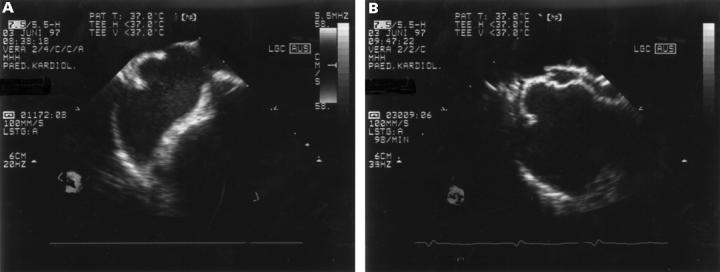
Figure 2 .
Selected video frame from transoesophageal echocardiographic study of patient 6 (transverse plane), in whom (A) lack of superior and anterior defect rim required (B) safe positioning of one right atrial arm of the device in the mouth of the superior caval vein before releasing the device.
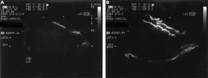
Figure 3 .
(A) Transoesophageal echocardiographic imaging of patient 7 (longitudinal plane) of the large inferiorly located septal defect and additionally smaller defect. (B) Imaging immediately after release of the second inferiorly located device. There was little overlapping of the distal arms of both devices.
Selected References
These references are in PubMed. This may not be the complete list of references from this article.
- Boutin C., Musewe N. N., Smallhorn J. F., Dyck J. D., Kobayashi T., Benson L. N. Echocardiographic follow-up of atrial septal defect after catheter closure by double-umbrella device. Circulation. 1993 Aug;88(2):621–627. doi: 10.1161/01.cir.88.2.621. [DOI] [PubMed] [Google Scholar]
- Bridges N. D., Hellenbrand W., Latson L., Filiano J., Newburger J. W., Lock J. E. Transcatheter closure of patent foramen ovale after presumed paradoxical embolism. Circulation. 1992 Dec;86(6):1902–1908. doi: 10.1161/01.cir.86.6.1902. [DOI] [PubMed] [Google Scholar]
- Das G. S., Voss G., Jarvis G., Wyche K., Gunther R., Wilson R. F. Experimental atrial septal defect closure with a new, transcatheter, self-centering device. Circulation. 1993 Oct;88(4 Pt 1):1754–1764. doi: 10.1161/01.cir.88.4.1754. [DOI] [PubMed] [Google Scholar]
- Ende D. J., Chopra P. S., Rao P. S. Transcatheter closure of atrial septal defect or patent foramen ovale with the buttoned device for prevention of recurrence of paradoxic embolism. Am J Cardiol. 1996 Jul 15;78(2):233–236. [PubMed] [Google Scholar]
- Ferreira S. M., Ho S. Y., Anderson R. H. Morphological study of defects of the atrial septum within the oval fossa: implications for transcatheter closure of left-to-right shunt. Br Heart J. 1992 Apr;67(4):316–320. doi: 10.1136/hrt.67.4.316. [DOI] [PMC free article] [PubMed] [Google Scholar]
- Godart F., Rey C., Francart C., Jarrar M., Vaksmann G. Two-dimensional echocardiographic and color Doppler measurements of atrial septal defect, and comparison with the balloon-stretched diameter. Am J Cardiol. 1993 Nov 1;72(14):1095–1097. doi: 10.1016/0002-9149(93)90873-b. [DOI] [PubMed] [Google Scholar]
- Hausdorf G., Schneider M., Franzbach B., Kampmann C., Kargus K., Goeldner B. Transcatheter closure of secundum atrial septal defects with the atrial septal defect occlusion system (ASDOS): initial experience in children. Heart. 1996 Jan;75(1):83–88. doi: 10.1136/hrt.75.1.83. [DOI] [PMC free article] [PubMed] [Google Scholar]
- Hickey P. R., Wessel D. L., Streitz S. L., Fox M. L., Kern F. H., Bridges N. D., Hansen D. D. Transcatheter closure of atrial septal defects: hemodynamic complications and anesthetic management. Anesth Analg. 1992 Jan;74(1):44–50. doi: 10.1213/00000539-199201000-00008. [DOI] [PubMed] [Google Scholar]
- Justo R. N., Nykanen D. G., Boutin C., McCrindle B. W., Freedom R. M., Benson L. N. Clinical impact of transcatheter closure of secundum atrial septal defects with the double umbrella device. Am J Cardiol. 1996 Apr 15;77(10):889–892. doi: 10.1016/S0002-9149(97)89192-8. [DOI] [PubMed] [Google Scholar]
- King T. D., Thompson S. L., Mills N. L. Measurement of atrial septal defect during cardiac catheterization. Experimental and clinical results. Am J Cardiol. 1978 Mar;41(3):537–542. doi: 10.1016/0002-9149(78)90012-7. [DOI] [PubMed] [Google Scholar]
- Koike K., Echigo S., Kumate M., Kobayashi T., Isoda T., Ishii M., Ishizawa A., Kamiya T., Kato H. [Transcatheter closure of atrial septal defect with a prototype clamshell septal umbrella: one year follow-up]. J Cardiol. 1994 Jan-Feb;24(1):53–60. [PubMed] [Google Scholar]
- Rao P. S., Langhough R. Relationship of echocardiographic, shunt flow, and angiographic size to the stretched diameter of the atrial septal defect. Am Heart J. 1991 Aug;122(2):505–508. doi: 10.1016/0002-8703(91)91008-b. [DOI] [PubMed] [Google Scholar]
- Rao P. S., Sideris E. B., Hausdorf G., Rey C., Lloyd T. R., Beekman R. H., Worms A. M., Bourlon F., Onorato E., Khalilullah M. International experience with secundum atrial septal defect occlusion by the buttoned device. Am Heart J. 1994 Nov;128(5):1022–1035. doi: 10.1016/0002-8703(94)90602-5. [DOI] [PubMed] [Google Scholar]
- Reddy S. C., Rao P. S., Ewenko J., Koscik R., Wilson A. D. Echocardiographic predictors of success of catheter closure of atrial septal defect with the buttoned device. Am Heart J. 1995 Jan;129(1):76–82. doi: 10.1016/0002-8703(95)90046-2. [DOI] [PubMed] [Google Scholar]
- Redington A. N., Rigby M. L. Transcatheter closure of interatrial communications with a modified umbrella device. Br Heart J. 1994 Oct;72(4):372–377. doi: 10.1136/hrt.72.4.372. [DOI] [PMC free article] [PubMed] [Google Scholar]
- Rocchini A. P. Transcatheter closure of atrial septal defects. Past, present, and future. Circulation. 1990 Sep;82(3):1044–1045. doi: 10.1161/01.cir.82.3.1044. [DOI] [PubMed] [Google Scholar]
- Rosenfeld H. M., van der Velde M. E., Sanders S. P., Colan S. D., Parness I. A., Lock J. E., Spevak P. J. Echocardiographic predictors of candidacy for successful transcatheter atrial septal defect closure. Cathet Cardiovasc Diagn. 1995 Jan;34(1):29–34. doi: 10.1002/ccd.1810340308. [DOI] [PubMed] [Google Scholar]
- Schenck M. H., Sterba R., Foreman C. K., Latson L. A. Improvement in noninvasive electrophysiologic findings in children after transcatheter atrial septal defect closure. Am J Cardiol. 1995 Oct 1;76(10):695–698. doi: 10.1016/s0002-9149(99)80199-4. [DOI] [PubMed] [Google Scholar]


