Abstract
Objective—To clarify the problems in angiographic diagnosis of major coronary arteries crossing the right ventricular outflow tract. Design—A retrospective study with clinicomorphological correlations to ascertain any aberrant coronary arteries and variations in distribution of the normal right coronary arterial branches. Setting—Tertiary referral centre. Subjects—36 necropsy specimens together with the aortograms and surgical reports from 130 patients with tetralogy of Fallot. Results—A preventricular branch was found in 19% of cases with tetralogy of Fallot, but in none of 13 normal hearts. Aberrant origin of the anterior interventricular coronary artery was found in 14% of the specimens. The combination of "laid back" and straight lateral views, when reviewed retrospectively, identified this anomaly correctly in nine of 16 patients, with these findings confirmed at surgery in seven patients. A major branch initially thought to cross the outflow tract was shown retrospectively to be an infundibular artery in six, with surgical confirmation in four. It was a preventricular branch in another patient. Conclusions—Using the laid back view alone, infundibular and preventricular branches may be mistaken for a major aberrant artery. A combination of laid back and straight lateral views is needed to avoid false positive diagnosis. Keywords: angiography; congenital heart defects; tetralogy of Fallot; paediatric cardiology
Full Text
The Full Text of this article is available as a PDF (265.6 KB).
Figure 1 .
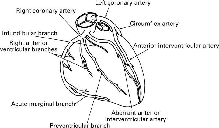
Relations of the various ventricular branches of the coronary arteries as seen on the sterno-costal surface of the heart. The course of the infundibular branch across the subpulmonary outflow tract distinguishes it from the right anterior ventricular branches. The preventricular branch is a prominent branch that passes diagonally across the anterior wall towards the apex. The aberrant anterior interventricular artery passes directly across the subpulmonary outflow tract to reach the interventricular groove and then runs toward the apex.
Figure 2 .
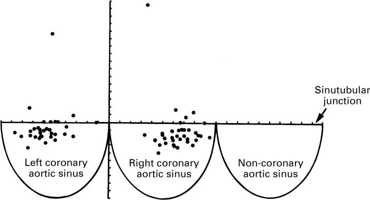
The location of the coronary orifices is plotted in relation to the depth of the sinuses and the distance from the zones of apposition between the facing leaflets. The horizontal and vertical markers represent 10% of the distances.
Figure 3 .
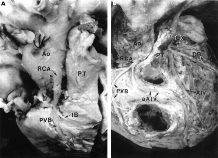
(A) The right coronary artery in this specimen arises well above the sinutubular junction. Its proximal course (between arrowheads) is through the aortic wall. The infundibular branch passes nearly horizontally across the distal right ventricular outflow tract, whereas the preventricular branch passes diagonally across the anterior wall. (B) An aberrant anterior interventricular artery arises from the right coronary artery in this specimen, which had had previous surgical repair. There is an accessory anterior interventricular artery from the left coronary artery, but this accessory artery terminates short of the apex. The preventricular branch is prominent. Ao, aorta; AIV, anterior interventricular artery; aAIV, aberrant anterior interventricular artery; Cx, circumflex coronary artery; DB, diagonal branch; IB, infundibular branch; PT, pulmonary trunk; PVB, preventricular branch; RCA, right coronary artery.
Figure 4 .
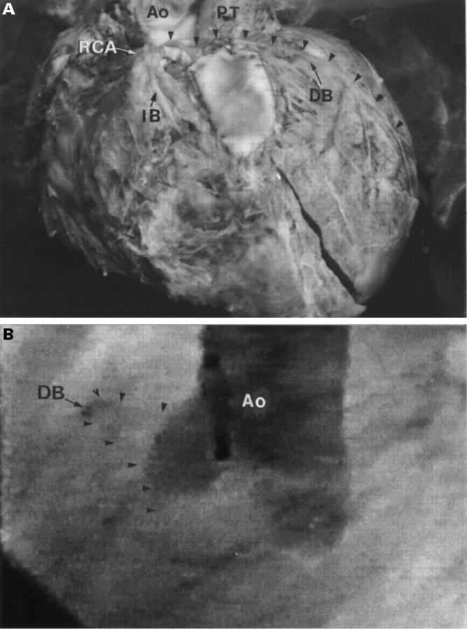
(A) The diagonal artery (arrowheads) passes across the subpulmonary infundibulum. It has been traumatised by sutures securing the outflow patch, resulting in infarction and thinning of the left ventricular wall. The anterior interventricular artery was absent. (B) The aberrant origin of the diagonal branch was not visualised on the atypical laid back view (case 15). Review of the straight lateral view shows the course of the aberrant artery (arrowheads). Ao, aorta; DB, diagonal branch; IB, infundibular branch; PT, pulmonary trunk; RCA, right coronary artery.
Figure 5 .
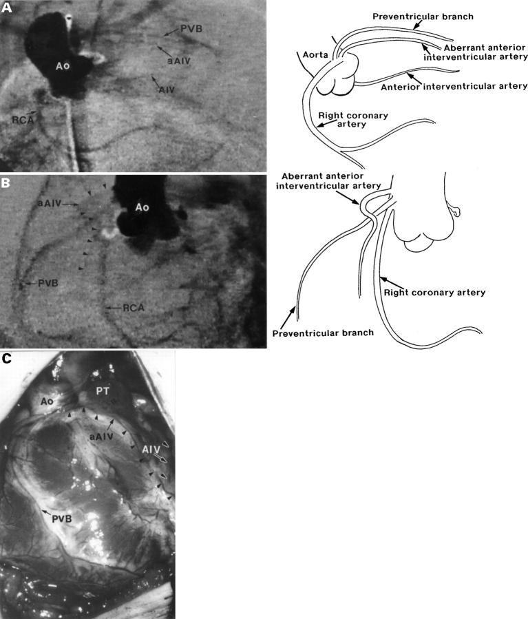
(A) This "laid back" view from case 3 shows two vessels in parallel, passing anterosuperiorly. (B) The straight lateral view separates these vessels into a large preventricular branch and a small aberrant anterior interventricular artery. (C) The intraoperative view confirms the findings. The aberrant anterior interventricular artery (black arrowheads) passes across the subpulmonary infundibulum, whereas the preventricular branch passes some distance away from the ventriculo-arterial junction. The left coronary artery also gives rise to an accessory anterior interventricular artery (black on white arrowheads). Ao, aorta; AIV, anterior interventricular artery; aAIV, aberrant anterior interventricular artery; PT, pulmonary trunk; PVB, preventricular branch; RCA, right coronary artery.
Figure 6 .
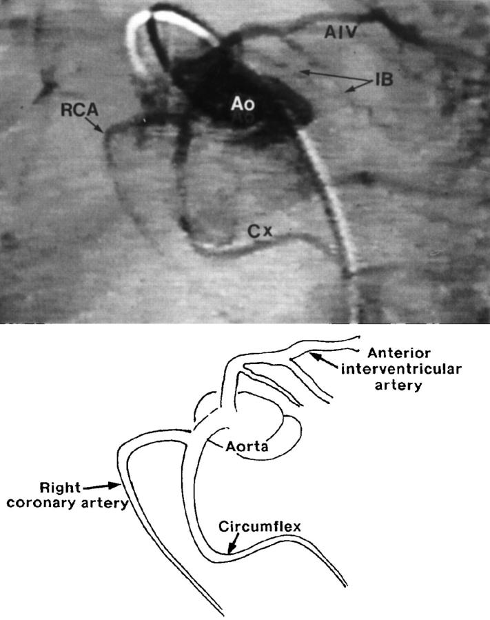
This "laid back" view from case 5 shows the branches from the single coronary artery. The diagram is composed from observations from a series of consecutive frames. The anterior interventricular artery passes from right to left across the subpulmonary infundibulum. Ao, aorta; AIV, anterior interventricular artery; Cx, circumflex coronary artery; IB, infundibular branch; RCA, right coronary artery.
Figure 7 .
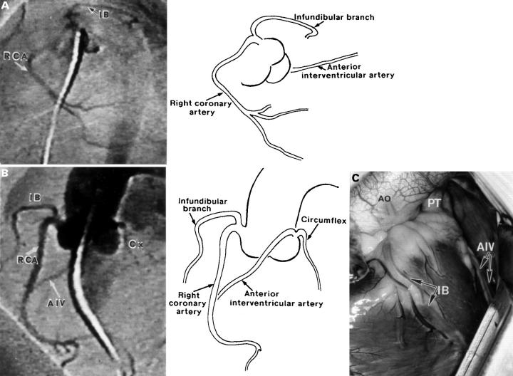
(A) The "laid back" view from case 8 gives the impression of an artery crossing antero-superiorly over the right ventricular outflow tract. (B) The straight lateral view clearly shows this to be the infundibular branch, distant from the normal anterior interventricular artery. (C) The intraoperative view confirms the findings. The infundibular branch crosses the subpulmonary infundibulum, and does not meet the normal anterior interventricular artery. Ao, aorta; AIV, anterior interventricular artery; IB, infundibular branch; PT, pulmonary trunk; RCA, right coronary artery.
Figure 8 .
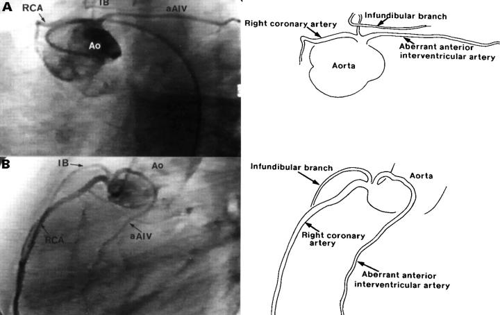
(A) The "laid back" view from case 16 shows two arteries passing antero-superiorly in a similar course. (B) The straight lateral view separates these two arteries into the infundibular branch and the aberrant anterior interventricular artery. The course of the aberrant artery was confirmed at surgery. In this projection, the infundibular branch passes forwards whereas the aberrant artery curves backwards and turns inferiorly. Ao, aorta; aAIV, aberrant anterior interventricular artery; IB, infundibular branch; RCA, right coronary artery.
Selected References
These references are in PubMed. This may not be the complete list of references from this article.
- Angelini P. Normal and anomalous coronary arteries: definitions and classification. Am Heart J. 1989 Feb;117(2):418–434. doi: 10.1016/0002-8703(89)90789-8. [DOI] [PubMed] [Google Scholar]
- Asou T., Karl T. R., Pawade A., Mee R. B. Arterial switch: translocation of the intramural coronary artery. Ann Thorac Surg. 1994 Feb;57(2):461–465. doi: 10.1016/0003-4975(94)91018-9. [DOI] [PubMed] [Google Scholar]
- Benson P. A. Anomalous aortic origin of coronary artery with sudden death: case report and review. Am Heart J. 1970 Feb;79(2):254–257. doi: 10.1016/0002-8703(70)90316-9. [DOI] [PubMed] [Google Scholar]
- Berquist T. H. Imaging of articular pathology: MRI, CT, arthrography. Clin Anat. 1997;10(1):1–13. doi: 10.1002/(SICI)1098-2353(1997)10:1<1::AID-CA1>3.0.CO;2-#. [DOI] [PubMed] [Google Scholar]
- Berry J. M., Jr, Einzig S., Krabill K. A., Bass J. L. Evaluation of coronary artery anatomy in patients with tetralogy of Fallot by two-dimensional echocardiography. Circulation. 1988 Jul;78(1):149–156. doi: 10.1161/01.cir.78.1.149. [DOI] [PubMed] [Google Scholar]
- Carvalho J. S., Silva C. M., Rigby M. L., Shinebourne E. A. Angiographic diagnosis of anomalous coronary artery in tetralogy of Fallot. Br Heart J. 1993 Jul;70(1):75–78. doi: 10.1136/hrt.70.1.75. [DOI] [PMC free article] [PubMed] [Google Scholar]
- Cheitlin M. D., De Castro C. M., McAllister H. A. Sudden death as a complication of anomalous left coronary origin from the anterior sinus of Valsalva, A not-so-minor congenital anomaly. Circulation. 1974 Oct;50(4):780–787. doi: 10.1161/01.cir.50.4.780. [DOI] [PubMed] [Google Scholar]
- Cohen L. S., Shaw L. D. Fatal myocardial infarction in an 11 year old boy associated with a unique coronary artery anomaly. Am J Cardiol. 1967 Mar;19(3):420–423. doi: 10.1016/0002-9149(67)90455-9. [DOI] [PubMed] [Google Scholar]
- Dabizzi R. P., Caprioli G., Aiazzi L., Castelli C., Baldrighi G., Parenzan L., Baldrighi V. Distribution and anomalies of coronary arteries in tetralogy of fallot. Circulation. 1980 Jan;61(1):95–102. doi: 10.1161/01.cir.61.1.95. [DOI] [PubMed] [Google Scholar]
- Dabizzi R. P., Teodori G., Barletta G. A., Caprioli G., Baldrighi G., Baldrighi V. Associated coronary and cardiac anomalies in the tetralogy of Fallot. An angiographic study. Eur Heart J. 1990 Aug;11(8):692–704. doi: 10.1093/oxfordjournals.eurheartj.a059784. [DOI] [PubMed] [Google Scholar]
- Fellows K. E., Freed M. D., Keane J. F., Praagh R., Bernhard W. F., Castaneda A. C. Results of routine preoperative coronary angiography in tetralogy of Fallot. Circulation. 1975 Mar;51(3):561–566. doi: 10.1161/01.cir.51.3.561. [DOI] [PubMed] [Google Scholar]
- Humes R. A., Driscoll D. J., Danielson G. K., Puga F. J. Tetralogy of Fallot with anomalous origin of left anterior descending coronary artery. Surgical options. J Thorac Cardiovasc Surg. 1987 Nov;94(5):784–787. [PubMed] [Google Scholar]
- Hurwitz R. A., Smith W., King H., Girod D. A., Caldwell R. L. Tetralogy of Fallot with abnormal coronary artery: 1967 to 1977. J Thorac Cardiovasc Surg. 1980 Jul;80(1):129–134. [PubMed] [Google Scholar]
- Jureidini S. B., Appleton R. S., Nouri S. Detection of coronary artery abnormalities in tetralogy of Fallot by two-dimensional echocardiography. J Am Coll Cardiol. 1989 Oct;14(4):960–967. doi: 10.1016/0735-1097(89)90473-7. [DOI] [PubMed] [Google Scholar]
- Kaushal S. K., Iyer K. S., Sharma R., Airan B., Bhan A., Das B., Saxena A., Venugopal P. Surgical experience with total correction of tetralogy of Fallot in infancy. Int J Cardiol. 1996 Sep;56(1):35–40. doi: 10.1016/0167-5273(96)02736-2. [DOI] [PubMed] [Google Scholar]
- LONGENECKER C. G., REEMTSMA K., CREECH O., Jr Anomalous coronary artery distribution associated with tetralogy of Fallot: a hazard in open cardiac repair. J Thorac Cardiovasc Surg. 1961 Aug;42:258–262. [PubMed] [Google Scholar]
- MENG C. C., ECKNER F. A., LEV M. CORONARY ARTERY DISTRIBUTION IN TETRALOGY OF FALLOT. Arch Surg. 1965 Mar;90:363–366. doi: 10.1001/archsurg.1965.01320090041009. [DOI] [PubMed] [Google Scholar]
- Mandell V. S., Lock J. E., Mayer J. E., Parness I. A., Kulik T. J. The "laid-back" aortogram: an improved angiographic view for demonstration of coronary arteries in transposition of the great arteries. Am J Cardiol. 1990 Jun 1;65(20):1379–1383. doi: 10.1016/0002-9149(90)91331-y. [DOI] [PubMed] [Google Scholar]
- Mustafa I., Gula G., Radley-Smith R., Durrer S., Yacoub M. Anomalous origin of the left coronary artery from the anterior aortic sinus: a potential cause of sudden death. Anatomic characterization and surgical treatment. J Thorac Cardiovasc Surg. 1981 Aug;82(2):297–300. [PubMed] [Google Scholar]
- O'Sullivan J., Bain H., Hunter S., Wren C. End-on aortogram: improved identification of important coronary artery anomalies in tetralogy of Fallot. Br Heart J. 1994 Jan;71(1):102–106. doi: 10.1136/hrt.71.1.102. [DOI] [PMC free article] [PubMed] [Google Scholar]
- Pasquini L., Parness I. A., Colan S. D., Wernovsky G., Mayer J. E., Sanders S. P. Diagnosis of intramural coronary artery in transposition of the great arteries using two-dimensional echocardiography. Circulation. 1993 Sep;88(3):1136–1141. doi: 10.1161/01.cir.88.3.1136. [DOI] [PubMed] [Google Scholar]
- Santoro G., Marino B., Di Carlo D., Formigari R., de Zorzi A., Mazzera E., Rinelli G., Marcelletti C., De Simone G., Pasquini L. Echocardiographically guided repair of tetralogy of Fallot. Am J Cardiol. 1994 Apr 15;73(11):808–811. doi: 10.1016/0002-9149(94)90885-0. [DOI] [PubMed] [Google Scholar]
- Shrivastava S., Edwards J. E. Coronary arterial origin in persistent truncus arteriosus. Circulation. 1977 Mar;55(3):551–554. doi: 10.1161/01.cir.55.3.551. [DOI] [PubMed] [Google Scholar]
- Shrivastava S., Mohan J. C., Mukhopadhyay S., Rajani M., Tandon R. Coronary artery anomalies in tetralogy of Fallot. Cardiovasc Intervent Radiol. 1987;10(4):215–218. doi: 10.1007/BF02593873. [DOI] [PubMed] [Google Scholar]
- Sim E. K., van Son J. A., Julsrud P. R., Puga F. J. Aortic intramural course of the left coronary artery in dextro-transposition of the great arteries. Ann Thorac Surg. 1994 Feb;57(2):458–460. doi: 10.1016/0003-4975(94)91017-0. [DOI] [PubMed] [Google Scholar]
- Stein P. D., Sabbah H. N. Orifice-view roentgenography for evaluation of the aortic valve. Am J Roentgenol Radium Ther Nucl Med. 1975 Dec;125(4):847–853. doi: 10.2214/ajr.125.4.847. [DOI] [PubMed] [Google Scholar]


