Abstract
A 44 year old man with Brugada syndrome and ventricular fibrillation had an autonomic disorder, shown by spectral analysis of heart rate variability and 123I-MIBG myocardial scintigraphy. Periodic variation of the ST segments was detected by Holter ECG. Increased high frequency power (0.15-0.40 Hz), an index of parasympathetic nerve activity, was observed just before ST segment elevation. In addition, local dysfunction of sympathetic nerves in the left ventricle was detected by 123I-MIBG myocardial scintigraphy. Unbalanced autonomic nerve function plays an important role in inducing Brugada-type ECG signs. Keywords: Brugada syndrome; autonomic disorder; 123I-MIBG myocardial scintigraphy
Full Text
The Full Text of this article is available as a PDF (91.7 KB).
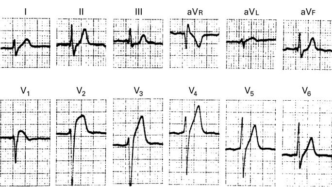
Figure 1 .
Standard 12 lead ECG on admission.
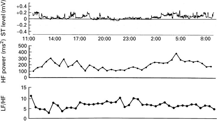
Figure 2 .
ST trendogram of Holter ECG and high frequency power obtained by analysis of spectral heart rate variability. ST segment elevation was observed in ST trendogram at 16:00 and 15:00 to 16:00. Before elevation of the ST segment, an increase in high frequency power, an index of parasympathetic nerve tension, was observed.
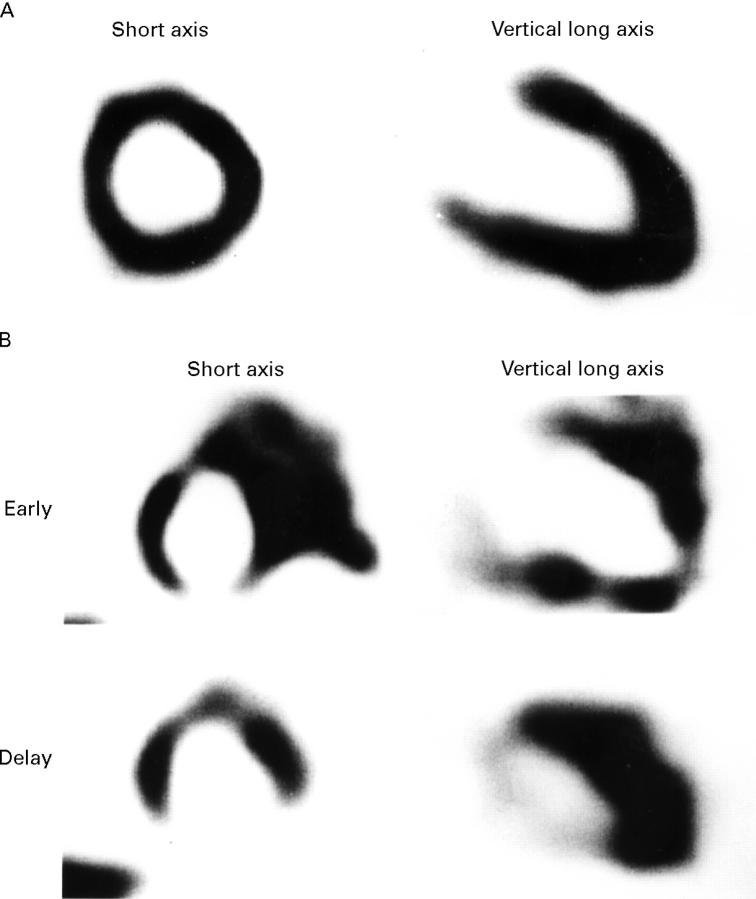
Figure 3 .
Myocardial scintigram using 99mTc-MIBI (A) or 123I-MIBG (B). In 99mTc-MIBI myocardial scintigraphy, no abnormality was observed in myocardial blood flow; however, during early imaging of myocardial scintigraphy using 123I-MIBG, a decreased accumulation or an unequal distribution of 123I-MIBG was observed in part of the inferior wall, the apex, and anterior wall of the left ventricle; while in delay imaging, an increased wash out of 123I-MIBG was observed in the inferior wall.


