Abstract
Acute ventricular septal rupture following myocardial infarction carries a high mortality. Early surgery improves survival but long term outcome depends on residual shunting and left ventricular function. Residual shunting is common despite apparently successful closure and may require reoperation. Transcatheter closure is an established method of treating selected congenital defects but clinical experience of transcatheter closure in postinfarction ventricular septal rupture is minimal. Transcatheter closure of a residual ventricular septal defect was successfully done using a new device, the Amplatzer septal occluder, in a 50 year old Indian man who had previously undergone emergency surgical repair for postinfarction acute ventricular septal rupture. The technique is described and its potential as a treatment in postinfarction ventricular septal rupture, its possible complications, and the important aspects of case selection and device design are discussed. Keywords: ventricular septal defect; transcatheter closure; Amplazter septal occluder
Full Text
The Full Text of this article is available as a PDF (102.5 KB).
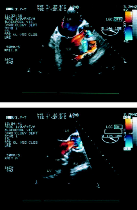
Figure 1 .

(Top) Preprocedure transoesophageal echocardiogram. Colour flow Doppler shows a significant left to right shunt across the VSD (arrow). (Bottom) Transoesophageal echocardiogram following deployment of the Amplatzer septal occluder (arrow) with obliteration of the left to right shunt. LA, left atrium; LV, left ventricle; RV, right ventricle; Ao, aorta.
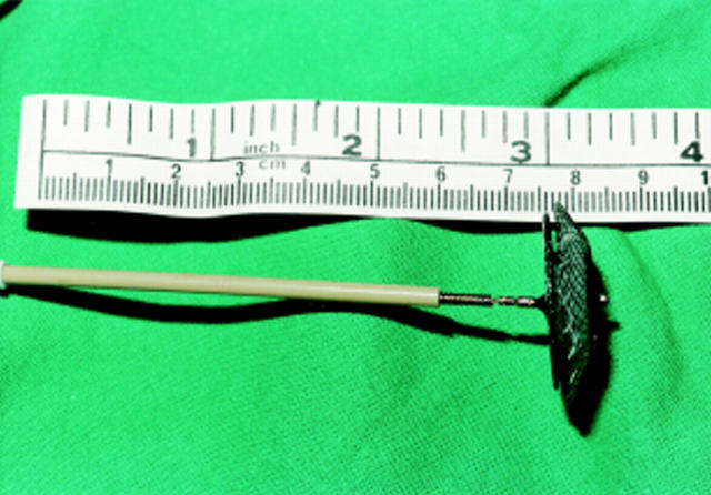
Figure 2 .
An Amplatzer septal occluder.
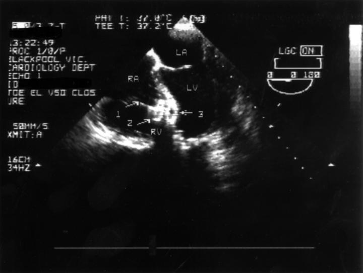
Figure 3 .
Transoesophageal echocardiogram showing the Amplatzer septal occluder positioned across the VSD before release from the delivery wire. LA, left atrium; LV, left ventricle; RA, right atrium; RV, right ventricle; arrow 1, delivery wire; arrow 2, proximal (right ventricular) disc; arrow 3, distal (left ventricular) disc.


