

Full Text
The Full Text of this article is available as a PDF (208.7 KB).

Figure 1 .

Typical card of information kept and written by Wood. It shows the correction of diagnosis after a second catheter on Angela Middleton who had the first cardiac catheterisation performed in the Brompton by Paul Wood and Walter Somerville.
Figure 2 .

Amalgam of the qualities of Paul Wood.
Figure 3 .

Annotation of physical signs in the case notes (1995) by senior registrar in paediatric cardiology (now a consultant). Boxed signs (arrow) show correct physical signs which indicate severe pulmonary stenosis NOT ventricular septal defect as suggested. Patient sent to surgery without catheter. Diagnosis confirmed.
Figure 4 .

Wood constructed simple figures from his data on cards, grading everything. This demonstrates the relation of effort intolerance (grade 1-4, with 4 the worst) to age of patients with secundum atrial septal defect.
Figure 5 .

Wood taking physiological investigation to the operating theatre. This shows the arterial pressure pulse when the finger was in the mitral valve (during mitral valvotomy) and when it is taken out following the valvotomy when the brachial pressure rises significantly.
Figure 6 .
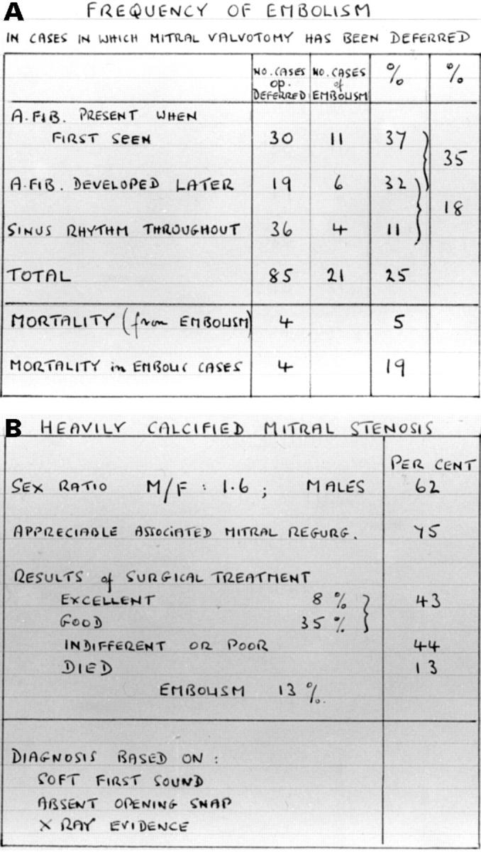
(A) Frequency of embolism in mitral stenosis. (B) Results of surgery with heavily calcified mitral stenosis. Nice illustration of Paul Wood's obsession with accuracy shown by comment on basis of diagnosis.
Figure 7 .

Paul Wood in characteristic pose on the left of Maurice Sokolow with Russell Brock on the right. Taken in the late 1950s.
Figure 8 .

The "music" (auscultation) of atrial septal defect (ASD) written by Paul Wood.
Figure 9 .

Paul Wood often described new disease in his letters to referring doctors. These letters show the description of idiopathic muscular subaortic stenosis and its physiology. (A) From Dr Richard Emanuel. (B) From Professors W McKenna and C Oakley.
Selected References
These references are in PubMed. This may not be the complete list of references from this article.
- GASUL B. M., DILLON R. F., VRLA V., HAIT G. Ventricular septal defects; their natural transformation into the cyanotic or noncyanotic type of tetralogy of Fallot. J Am Med Assoc. 1957 Jun 22;164(8):847–853. doi: 10.1001/jama.1957.02980080017003. [DOI] [PubMed] [Google Scholar]
- LOWN B., NEUMAN J., AMARASINGHAM R., BERKOVITS B. V. Comparison of alternating current with direct electroshock across the closed chest. Am J Cardiol. 1962 Aug;10:223–233. doi: 10.1016/0002-9149(62)90299-0. [DOI] [PubMed] [Google Scholar]
- Rabinovitch M., Haworth S. G., Castaneda A. R., Nadas A. S., Reid L. M. Lung biopsy in congenital heart disease: a morphometric approach to pulmonary vascular disease. Circulation. 1978 Dec;58(6):1107–1122. doi: 10.1161/01.cir.58.6.1107. [DOI] [PubMed] [Google Scholar]
- Somerville J. Congenital heart disease--changes in form and function. Br Heart J. 1979 Jan;41(1):1–22. doi: 10.1136/hrt.41.1.1. [DOI] [PMC free article] [PubMed] [Google Scholar]
- WOOD P. The Eisenmenger syndrome or pulmonary hypertension with reversed central shunt. I. Br Med J. 1958 Sep 20;2(5098):701–709. doi: 10.1136/bmj.2.5098.701. [DOI] [PMC free article] [PubMed] [Google Scholar]