Full Text
The Full Text of this article is available as a PDF (95.6 KB).
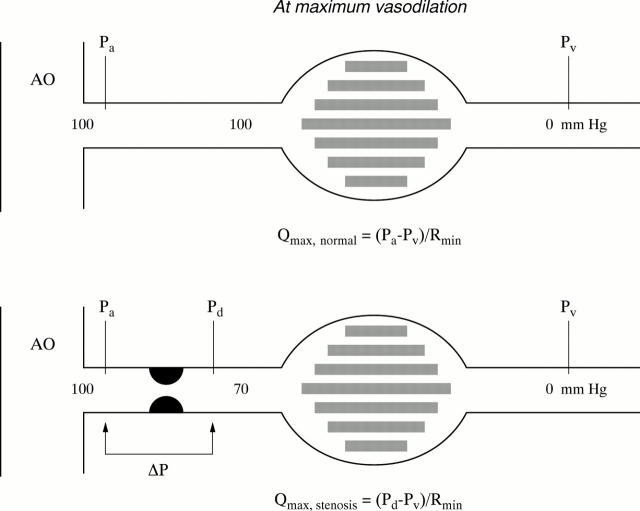
Figure 1 .
Schematic illustration of the coronary artery and its dependent myocardial vascular bed. Myocardial blood flow equals the perfusion pressure across the myocardium, divided by myocardial resistance. Because at maximum arteriolar vasodilatation resistance is minimal and constant (Rmin), maximum flow in the stenotic situation as a ratio to normal maximum flow equals the ratio of the myocardial perfusion pressure in the presence of the stenosis (Pd − Pv) to normal myocardial perfusion pressure (Pa − Pv), both measured after administration of a maximum hyperaemic stimulus. In other words, FFR equals (Pd − Pv)/(Pa − Pv ), which is generally very close to Pd/Pa . In this example, FFR equals 0.70. AO, aorta; Pa, Pd, and Pv, mean aortic, distal coronary, and central venous pressure measured at maximum coronary hyperaemia; Qmax , normal, maximum achievable myocardial flow if the coronary artery were normal; Qmax, stenosis, maximum achievable myocardial flow in the presence of a stenosis; Rmin indicates minimal resistance of the myocardial vascular bed.
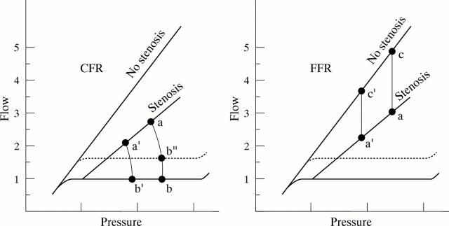
Figure 2 .
Features of coronary flow reserve (CFR) and fractional flow reserve (FFR). The coronary pressure-flow relation at maximum hyperaemia is indicated both for a normal coronary artery and a coronary stenosis. The resting pressure-flow relation (autoregulatory range) is represented by the solid horizontal line. (Left) CFR is defined as the ratio of hyperaemic to resting flow (a/b). It is obvious that with changing blood pressure, the value of CFR changes to a'/b'. Also, when resting flow changes (as with changing heart rate or contractility) CFR changes to a/b". Therefore, no uniform normal value of CFR can be defined. (Right) FFR is defined as the ratio of maximum flow in the presence of a stenosis divided by normal maximum flow (a/c). This ratio remains unaffected by changes in blood pressure (a/c = a'/c') or by variations in resting flow. Its normal value is always 1.0 , irrespective of the patient, coronary artery, or prevailing haemodynamic conditions. Therefore, FFR is a more specific measure of functional stenosis severity.
Selected References
These references are in PubMed. This may not be the complete list of references from this article.
- Bartunek J., Marwick T. H., Rodrigues A. C., Vincent M., Van Schuerbeeck E., Sys S. U., de Bruyne B. Dobutamine-induced wall motion abnormalities: correlations with myocardial fractional flow reserve and quantitative coronary angiography. J Am Coll Cardiol. 1996 May;27(6):1429–1436. doi: 10.1016/0735-1097(96)00022-8. [DOI] [PubMed] [Google Scholar]
- Bech G. J., De Bruyne B., Bonnier H. J., Bartunek J., Wijns W., Peels K., Heyndrickx G. R., Koolen J. J., Pijls N. H. Long-term follow-up after deferral of percutaneous transluminal coronary angioplasty of intermediate stenosis on the basis of coronary pressure measurement. J Am Coll Cardiol. 1998 Mar 15;31(4):841–847. doi: 10.1016/s0735-1097(98)00050-3. [DOI] [PubMed] [Google Scholar]
- De Bruyne B., Bartunek J., Sys S. U., Heyndrickx G. R. Relation between myocardial fractional flow reserve calculated from coronary pressure measurements and exercise-induced myocardial ischemia. Circulation. 1995 Jul 1;92(1):39–46. doi: 10.1161/01.cir.92.1.39. [DOI] [PubMed] [Google Scholar]
- De Bruyne B., Baudhuin T., Melin J. A., Pijls N. H., Sys S. U., Bol A., Paulus W. J., Heyndrickx G. R., Wijns W. Coronary flow reserve calculated from pressure measurements in humans. Validation with positron emission tomography. Circulation. 1994 Mar;89(3):1013–1022. doi: 10.1161/01.cir.89.3.1013. [DOI] [PubMed] [Google Scholar]
- De Bruyne B., Sys S. U., Heyndrickx G. R. Percutaneous transluminal coronary angioplasty catheters versus fluid-filled pressure monitoring guidewires for coronary pressure measurements and correlation with quantitative coronary angiography. Am J Cardiol. 1993 Nov 15;72(15):1101–1106. doi: 10.1016/0002-9149(93)90976-j. [DOI] [PubMed] [Google Scholar]
- Kern M. J., de Bruyne B., Pijls N. H. From research to clinical practice: current role of intracoronary physiologically based decision making in the cardiac catheterization laboratory. J Am Coll Cardiol. 1997 Sep;30(3):613–620. doi: 10.1016/s0735-1097(97)00224-6. [DOI] [PubMed] [Google Scholar]
- Pijls N. H., Bech G. J., el Gamal M. I., Bonnier H. J., De Bruyne B., Van Gelder B., Michels H. R., Koolen J. J. Quantification of recruitable coronary collateral blood flow in conscious humans and its potential to predict future ischemic events. J Am Coll Cardiol. 1995 Jun;25(7):1522–1528. doi: 10.1016/0735-1097(95)00111-g. [DOI] [PubMed] [Google Scholar]
- Pijls N. H., De Bruyne B., Peels K., Van Der Voort P. H., Bonnier H. J., Bartunek J Koolen J. J., Koolen J. J. Measurement of fractional flow reserve to assess the functional severity of coronary-artery stenoses. N Engl J Med. 1996 Jun 27;334(26):1703–1708. doi: 10.1056/NEJM199606273342604. [DOI] [PubMed] [Google Scholar]
- Pijls N. H., Van Gelder B., Van der Voort P., Peels K., Bracke F. A., Bonnier H. J., el Gamal M. I. Fractional flow reserve. A useful index to evaluate the influence of an epicardial coronary stenosis on myocardial blood flow. Circulation. 1995 Dec 1;92(11):3183–3193. doi: 10.1161/01.cir.92.11.3183. [DOI] [PubMed] [Google Scholar]
- Pijls N. H., van Son J. A., Kirkeeide R. L., De Bruyne B., Gould K. L. Experimental basis of determining maximum coronary, myocardial, and collateral blood flow by pressure measurements for assessing functional stenosis severity before and after percutaneous transluminal coronary angioplasty. Circulation. 1993 Apr;87(4):1354–1367. doi: 10.1161/01.cir.87.4.1354. [DOI] [PubMed] [Google Scholar]
- Topol E. J., Nissen S. E. Our preoccupation with coronary luminology. The dissociation between clinical and angiographic findings in ischemic heart disease. Circulation. 1995 Oct 15;92(8):2333–2342. doi: 10.1161/01.cir.92.8.2333. [DOI] [PubMed] [Google Scholar]
- Wilson R. F. Assessing the severity of coronary-artery stenoses. N Engl J Med. 1996 Jun 27;334(26):1735–1737. doi: 10.1056/NEJM199606273342610. [DOI] [PubMed] [Google Scholar]
- de Bruyne B., Bartunek J., Sys S. U., Pijls N. H., Heyndrickx G. R., Wijns W. Simultaneous coronary pressure and flow velocity measurements in humans. Feasibility, reproducibility, and hemodynamic dependence of coronary flow velocity reserve, hyperemic flow versus pressure slope index, and fractional flow reserve. Circulation. 1996 Oct 15;94(8):1842–1849. doi: 10.1161/01.cir.94.8.1842. [DOI] [PubMed] [Google Scholar]


