Abstract
Objective—To examine the relation between the initial microvascular perfusion pattern, as assessed by intracoronary myocardial contrast echocardiography (MCE), immediately after restoration of TIMI (thrombolysis in myocardial infarction) (TIMI) grade 3 flow during acute myocardial infarction, and the extent and timing of functional recovery in the area at risk. Setting—Referral centre for interventional cardiology. Methods—Intracoronary MCE was performed 15 minutes after TIMI grade 3 recanalisation of the infarct artery in 25 patients. Segmental myocardial contrast patterns were graded semiquantitatively (0, none; 0.5, heterogeneous; 1, homogeneous). Functional recovery was assessed by echocardiography on days 9 and 42. Results—Among 174 myocardial segments in the area at risk, wall motion recovery on day 9 was observed in 40% of MCE grade 1 segments but there was no significant recovery in grade 0 or 0.5 segments. On day 42, recovery had occurred in 56% of MCE grade 1 segments (p < 0.0001 v MCE grade 0 and 0.5; p = 0.0001 v MCE grade 1 on day 9), and 22% of MCE grade 0.5 segments (p = 0.02 v MCE grade 0; p = 0.0005 v MCE grade 0.5 on day 9); MCE grade 0 segments did not recover. Negative predictive value in predicting recovery by contrast enhancement was 95% and 89% by days 9 and 42, respectively. Conclusions—Contractile recovery occurs earliest in well reperfused segments. Up to one quarter of segments with heterogeneous contrast enhancement show wall motion recovery within the first six weeks. Myocardial perfusion after recanalisation in acute myocardial infarction, even if heterogeneous, is a prerequisite for postischaemic functional recovery. Thus preservation of acute myocardial perfusion is associated with more complete and early functional recovery. Keywords: acute myocardial infarction; myocardial contrast echocardiography; microcirculation; functional recovery
Full Text
The Full Text of this article is available as a PDF (139.3 KB).
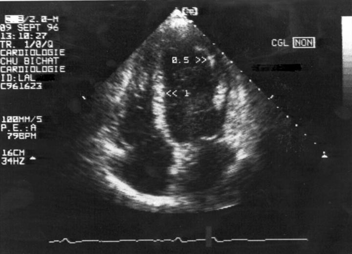
Figure 1 .
Apical four chamber view obtained by myocardial contrast echocardiography (MCE) in a patient immediately after successful primary angioplasty of an occluded left anterior descending coronary artery. The mid-septal segment is well perfused (MCE grade 1); the apicolateral segment shows only subendocardial perfusion (MCE grade 0.5).
Figure 2 .
Apical two chamber view of the same patient. The apical segment of the anterior wall has no tissue perfusion (MCE grade 0), whereas the mid-segment of the inferior wall is well perfused (MCE grade 1).
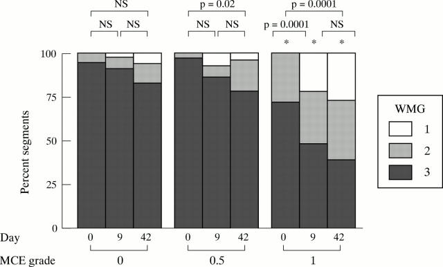
Figure 3 .
Bar graph showing serial echocardiographic segmental wall motion assessment (at day 0, day 9, and day 42) as a function of acute (day 0) myocardial contrast pattern. MCE, myocardial contrast echocardiography; WMG, wall motion grade. *p = 0.0001 v perfusion grade 0 and 0.5 on the same day
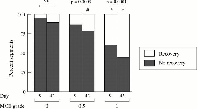
Figure 4 .
Bar graph showing segmental wall motion recovery (on day 9 and day 42) as a function of acute (day 0) myocardial contrast pattern. For each segment, recovery was defined as an improvement of at least one grade in the wall motion score from day 0 to day 9 or from day 0 to day 42. MCE, myocardial contrast echocardiography. *p < 0.05 v contrast grade 0 and 0.5 the same day; #p < 0.05 v contrast grade 0 the same day.
Selected References
These references are in PubMed. This may not be the complete list of references from this article.
- Agati L., Voci P., Autore C., Luongo R., Testa G., Mallus M. T., Di Roma A., Fedele F., Dagianti A. Combined use of dobutamine echocardiography and myocardial contrast echocardiography in predicting regional dysfunction recovery after coronary revascularization in patients with recent myocardial infarction. Eur Heart J. 1997 May;18(5):771–779. doi: 10.1093/oxfordjournals.eurheartj.a015342. [DOI] [PubMed] [Google Scholar]
- Agati L., Voci P., Bilotta F., Luongo R., Autore C., Penco M., Iacoboni C., Fedele F., Dagianti A. Influence of residual perfusion within the infarct zone on the natural history of left ventricular dysfunction after acute myocardial infarction: a myocardial contrast echocardiographic study. J Am Coll Cardiol. 1994 Aug;24(2):336–342. doi: 10.1016/0735-1097(94)90285-2. [DOI] [PubMed] [Google Scholar]
- Anderson J. L., Karagounis L. A., Becker L. C., Sorensen S. G., Menlove R. L. TIMI perfusion grade 3 but not grade 2 results in improved outcome after thrombolysis for myocardial infarction. Ventriculographic, enzymatic, and electrocardiographic evidence from the TEAM-3 Study. Circulation. 1993 Jun;87(6):1829–1839. doi: 10.1161/01.cir.87.6.1829. [DOI] [PubMed] [Google Scholar]
- Bodenheimer M. M., Banka V. S., Hermann G. A., Trout R. G., Pasdar H., Helfant R. H. Reversible asynergy. Histopathologic and electrographic correlations in patients with coronary artery disease. Circulation. 1976 May;53(5):792–796. doi: 10.1161/01.cir.53.5.792. [DOI] [PubMed] [Google Scholar]
- Bolli R. Myocardial 'stunning' in man. Circulation. 1992 Dec;86(6):1671–1691. doi: 10.1161/01.cir.86.6.1671. [DOI] [PubMed] [Google Scholar]
- Bolognese L., Antoniucci D., Rovai D., Buonamici P., Cerisano G., Santoro G. M., Marini C., L'Abbate A., Fazzini P. F. Myocardial contrast echocardiography versus dobutamine echocardiography for predicting functional recovery after acute myocardial infarction treated with primary coronary angioplasty. J Am Coll Cardiol. 1996 Dec;28(7):1677–1683. doi: 10.1016/S0735-1097(96)00400-7. [DOI] [PubMed] [Google Scholar]
- Braunwald E., Kloner R. A. The stunned myocardium: prolonged, postischemic ventricular dysfunction. Circulation. 1982 Dec;66(6):1146–1149. doi: 10.1161/01.cir.66.6.1146. [DOI] [PubMed] [Google Scholar]
- Chesebro J. H., Knatterud G., Roberts R., Borer J., Cohen L. S., Dalen J., Dodge H. T., Francis C. K., Hillis D., Ludbrook P. Thrombolysis in Myocardial Infarction (TIMI) Trial, Phase I: A comparison between intravenous tissue plasminogen activator and intravenous streptokinase. Clinical findings through hospital discharge. Circulation. 1987 Jul;76(1):142–154. doi: 10.1161/01.cir.76.1.142. [DOI] [PubMed] [Google Scholar]
- Costanzo M. R., Augustine S., Bourge R., Bristow M., O'Connell J. B., Driscoll D., Rose E. Selection and treatment of candidates for heart transplantation. A statement for health professionals from the Committee on Heart Failure and Cardiac Transplantation of the Council on Clinical Cardiology, American Heart Association. Circulation. 1995 Dec 15;92(12):3593–3612. doi: 10.1161/01.cir.92.12.3593. [DOI] [PubMed] [Google Scholar]
- Force T., Kemper A., Perkins L., Gilfoil M., Cohen C., Parisi A. F. Overestimation of infarct size by quantitative two-dimensional echocardiography: the role of tethering and of analytic procedures. Circulation. 1986 Jun;73(6):1360–1368. doi: 10.1161/01.cir.73.6.1360. [DOI] [PubMed] [Google Scholar]
- Galli M., Marcassa C., Bolli R., Giannuzzi P., Temporelli P. L., Imparato A., Silva Orrego P. L., Giubbini R., Giordano A., Tavazzi L. Spontaneous delayed recovery of perfusion and contraction after the first 5 weeks after anterior infarction. Evidence for the presence of hibernating myocardium in the infarcted area. Circulation. 1994 Sep;90(3):1386–1397. doi: 10.1161/01.cir.90.3.1386. [DOI] [PubMed] [Google Scholar]
- Iliceto S., Galiuto L., Marchese A., Cavallari D., Colonna P., Biasco G., Rizzon P. Analysis of microvascular integrity, contractile reserve, and myocardial viability after acute myocardial infarction by dobutamine echocardiography and myocardial contrast echocardiography. Am J Cardiol. 1996 Mar 1;77(7):441–445. doi: 10.1016/s0002-9149(97)89334-4. [DOI] [PubMed] [Google Scholar]
- Ito H., Iwakura K., Oh H., Masuyama T., Hori M., Higashino Y., Fujii K., Minamino T. Temporal changes in myocardial perfusion patterns in patients with reperfused anterior wall myocardial infarction. Their relation to myocardial viability. Circulation. 1995 Feb 1;91(3):656–662. doi: 10.1161/01.cir.91.3.656. [DOI] [PubMed] [Google Scholar]
- Ito H., Maruyama A., Iwakura K., Takiuchi S., Masuyama T., Hori M., Higashino Y., Fujii K., Minamino T. Clinical implications of the 'no reflow' phenomenon. A predictor of complications and left ventricular remodeling in reperfused anterior wall myocardial infarction. Circulation. 1996 Jan 15;93(2):223–228. doi: 10.1161/01.cir.93.2.223. [DOI] [PubMed] [Google Scholar]
- Ito H., Okamura A., Iwakura K., Masuyama T., Hori M., Takiuchi S., Negoro S., Nakatsuchi Y., Taniyama Y., Higashino Y. Myocardial perfusion patterns related to thrombolysis in myocardial infarction perfusion grades after coronary angioplasty in patients with acute anterior wall myocardial infarction. Circulation. 1996 Jun 1;93(11):1993–1999. doi: 10.1161/01.cir.93.11.1993. [DOI] [PubMed] [Google Scholar]
- Ito H., Tomooka T., Sakai N., Higashino Y., Fujii K., Katoh O., Masuyama T., Kitabatake A., Minamino T. Time course of functional improvement in stunned myocardium in risk area in patients with reperfused anterior infarction. Circulation. 1993 Feb;87(2):355–362. doi: 10.1161/01.cir.87.2.355. [DOI] [PubMed] [Google Scholar]
- Ito H., Tomooka T., Sakai N., Yu H., Higashino Y., Fujii K., Masuyama T., Kitabatake A., Minamino T. Lack of myocardial perfusion immediately after successful thrombolysis. A predictor of poor recovery of left ventricular function in anterior myocardial infarction. Circulation. 1992 May;85(5):1699–1705. doi: 10.1161/01.cir.85.5.1699. [DOI] [PubMed] [Google Scholar]
- Kaul S., Senior R., Dittrich H., Raval U., Khattar R., Lahiri A. Detection of coronary artery disease with myocardial contrast echocardiography: comparison with 99mTc-sestamibi single-photon emission computed tomography. Circulation. 1997 Aug 5;96(3):785–792. [PubMed] [Google Scholar]
- Myers J. H., Stirling M. C., Choy M., Buda A. J., Gallagher K. P. Direct measurement of inner and outer wall thickening dynamics with epicardial echocardiography. Circulation. 1986 Jul;74(1):164–172. doi: 10.1161/01.cir.74.1.164. [DOI] [PubMed] [Google Scholar]
- Porter T. R., Li S., Kricsfeld D., Armbruster R. W. Detection of myocardial perfusion in multiple echocardiographic windows with one intravenous injection of microbubbles using transient response second harmonic imaging. J Am Coll Cardiol. 1997 Mar 15;29(4):791–799. doi: 10.1016/s0735-1097(96)00575-x. [DOI] [PubMed] [Google Scholar]
- Ragosta M., Camarano G., Kaul S., Powers E. R., Sarembock I. J., Gimple L. W. Microvascular integrity indicates myocellular viability in patients with recent myocardial infarction. New insights using myocardial contrast echocardiography. Circulation. 1994 Jun;89(6):2562–2569. doi: 10.1161/01.cir.89.6.2562. [DOI] [PubMed] [Google Scholar]
- Sabia P. J., Powers E. R., Jayaweera A. R., Ragosta M., Kaul S. Functional significance of collateral blood flow in patients with recent acute myocardial infarction. A study using myocardial contrast echocardiography. Circulation. 1992 Jun;85(6):2080–2089. doi: 10.1161/01.cir.85.6.2080. [DOI] [PubMed] [Google Scholar]
- Schiller N. B., Shah P. M., Crawford M., DeMaria A., Devereux R., Feigenbaum H., Gutgesell H., Reichek N., Sahn D., Schnittger I. Recommendations for quantitation of the left ventricle by two-dimensional echocardiography. American Society of Echocardiography Committee on Standards, Subcommittee on Quantitation of Two-Dimensional Echocardiograms. J Am Soc Echocardiogr. 1989 Sep-Oct;2(5):358–367. doi: 10.1016/s0894-7317(89)80014-8. [DOI] [PubMed] [Google Scholar]
- Touchstone D. A., Beller G. A., Nygaard T. W., Tedesco C., Kaul S. Effects of successful intravenous reperfusion therapy on regional myocardial function and geometry in humans: a tomographic assessment using two-dimensional echocardiography. J Am Coll Cardiol. 1989 Jun;13(7):1506–1513. doi: 10.1016/0735-1097(89)90340-9. [DOI] [PubMed] [Google Scholar]
- Villanueva F. S., Glasheen W. P., Sklenar J., Kaul S. Assessment of risk area during coronary occlusion and infarct size after reperfusion with myocardial contrast echocardiography using left and right atrial injections of contrast. Circulation. 1993 Aug;88(2):596–604. doi: 10.1161/01.cir.88.2.596. [DOI] [PubMed] [Google Scholar]
- Weintraub W. S., Hattori S., Agarwal J. B., Bodenheimer M. M., Banka V. S., Helfant R. H. The relationship between myocardial blood flow and contraction by myocardial layer in the canine left ventricle during ischemia. Circ Res. 1981 Mar;48(3):430–438. doi: 10.1161/01.res.48.3.430. [DOI] [PubMed] [Google Scholar]


