Abstract
Takayasu aortitis is a chronic inflammatory vasculitis characterised by stenosis or obliteration of large and medium sized arteries. Although coronary arteries are affected in approximately 10% of cases, most of the lesions are luminal narrowing, and coronary aneurysm formation is extremely rare. A case is described of giant aneurysm of the left main coronary artery complicated with Takayasu aortitis in a 46 year old Japanese woman who was followed until her death at age 71. Pronounced intimal proliferation and adventitial fibrous thickening of the involved arterial wall usually induce constriction or occlusion at the orifice of the main branch of the aorta in Takayasu aortitis. However, systemic hypertension, which resulted from renovascular stenoses in this case, is likely to have enlarged the vessel lumen before replacement of medial and adventitial fibrosis after extensive destruction of medial elastic fibres in the left main coronary artery. Moreover, associations such as autoimmune hepatitis, chronic thyroiditis, and Sjögren syndrome strongly suggests that Takayasu aortitis may be an autoimmune disease. Keywords: giant coronary aneurysm; Takayasu aortitis; renovascular hypertension; autoimmune disorders
Full Text
The Full Text of this article is available as a PDF (124.8 KB).
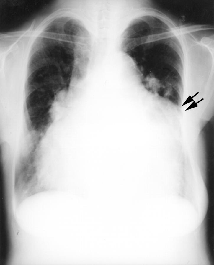
Figure 1 .
Severe cardiac enlargement and a prominent bulge (arrows) on the third arch of the left heart border.
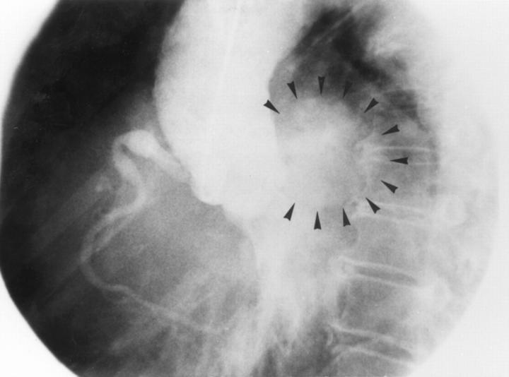
Figure 2 .
Left anterior cranial view of ascending aortogram at diastolic phase. A root aortogram shows dilatation of ascending aorta, severe aortic regurgitation, saccular or oval giant coronary aneurysm (surrounded by arrows) of left main stem, and small aneurysm of right coronary artery.
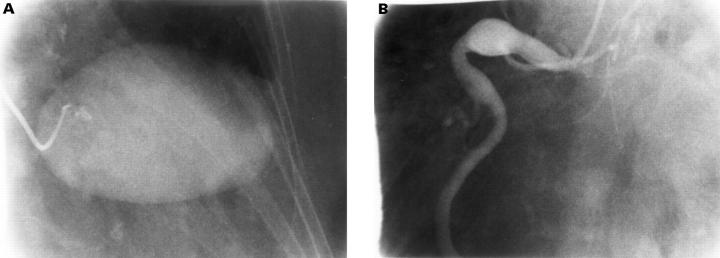
Figure 3 .
(A) Coronary angiogram showing saccular giant aneurysm of the left main coronary artery. (B) Coronary angiogram in the left anterior oblique showing an enlarged fusiform aneurysm in the proximal segment of the right coronary artery.
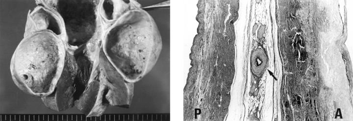
Figure 4 .
(Left) Massive aneurysm of left main coronary artery with its thickened wall and atherosclerotic change. (Right) Severely scarred media is surrounded by dense adventitial and atherosclerotic intima in the ascending aortic wall (A) and the pulmonary arterial wall (P). A vasa vasorum (arrow) is characteristically narrowed. (Elastic van Gieson stain, original magnification ×60.)


