

Abstract
Objective—To study patients with atrioventricular septal defect to determine the pathognomonic morphological features of the lesion and the relation between the septal structures and the atrioventricular junction. Setting —Tertiary level paediatric cardiology centre. Methods—Cross sectional echocardiograms from 60 patients were reviewed using qualitative and quantitative analysis. The unifying feature was the presence of a common atrioventricular junction. The overall dimensions of the septal defect were determined and related to the plane of the common junction; the extent of both the atrial and the ventricular septal components was then measured according to the site of closure of the bridging leaflets. Results—In 48 cases, the common junction was guarded by a common valvar orifice, but in 12 cases there were separate right and left valvar orifices. Irrespective of the valvar morphology, no significant difference was found between the groups in terms of the dimensions of the atrial and ventricular septal components. In all patients, the hole permitting shunting at atrial level extended below the plane of the atrioventricular junction, with a variable position of the leading edge of the atrial septum itself. Conclusions—The atrioventricular junction is a common structure irrespective of valvar morphology. In spite of the presence of unequivocal shunting at atrial level, the atrial septum is usually a well formed structure, even extending in some below the level of the common atrioventricular junction. Keywords: atrioventricular canal malformation; endocardial cushion defects; level of shunting; morphology
Full Text
The Full Text of this article is available as a PDF (219.8 KB).
Figure 1 .

Diagram showing the measurements taken. The plane of the atrioventricular junction is marked by the broken line (J), and is taken as the zero point in fig 4A and 4B. Note that in the right hand panel the lower edge of the atrial septum is below the level of the atrioventricular junction (S). A, lower edge of the atrial septum; B, atrial surface of the bridging leaflet; C, ventricular surface of the superior bridging leaflet; D, crest of the muscular septum.
Figure 2 .

The junctional arrangement of atrioventricular septal defect with common atrioventricular valve shown (A) echocardiographically and (B) in a postmortem specimen from a different patient. AO, aorta; BL, bridging leaflet; LV, left ventricle.
Figure 3 .
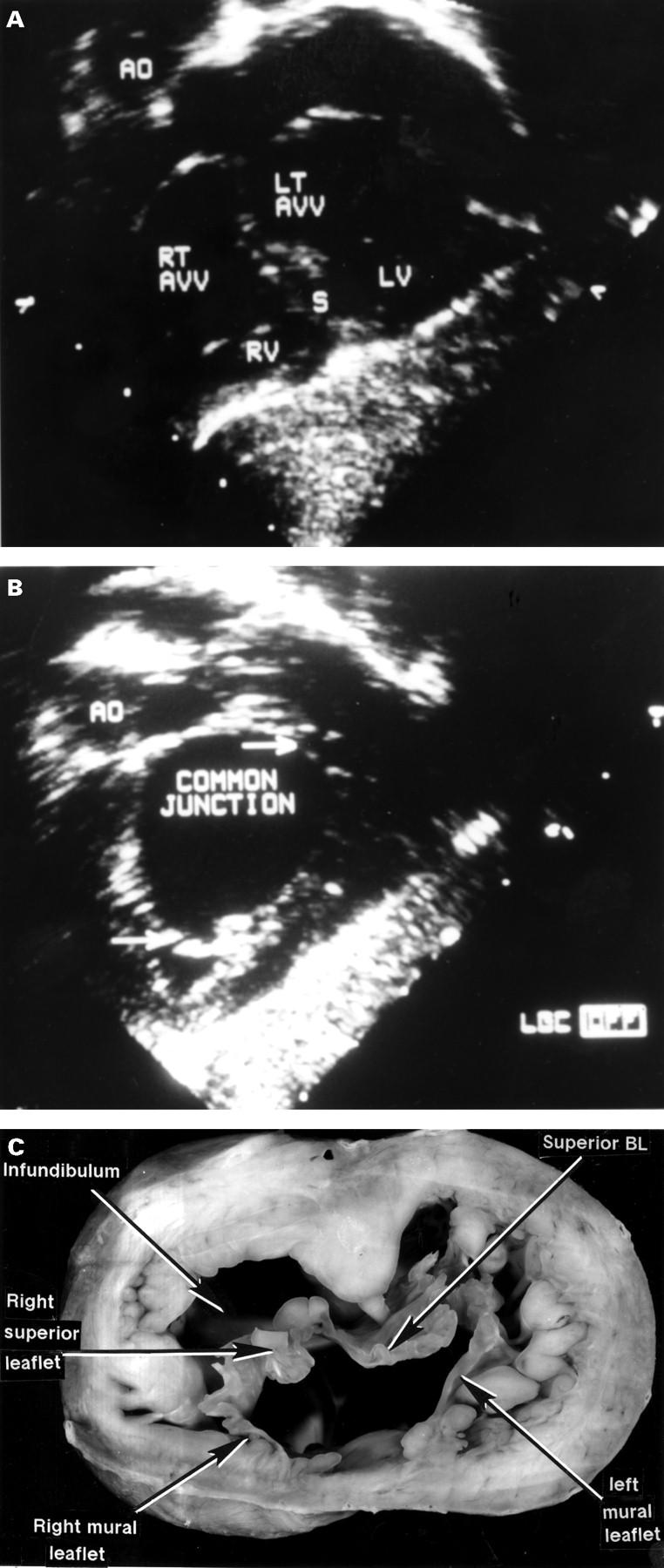
Comparable echocardiographic cuts (A, B) and a cross section from the heart of a different patient (C) showing the junctional arrangement in atrioventricular septal defect with common atrioventricular junction, but with separate valvar orifices for the right and left ventricles. The more distal echo cut (A) shows the common junction relative to the ventricular septum(s). As shown by the more proximal cut (B), at the level of the defect itself, the septum is not seen and the junction is obviously common (between arrows). This is confirmed by the cut of the postmortem specimen. AO, aorta; BL, bridging leaflet; LV, left ventricle; RT AVV, LI AVV, right and left atrioventricular valves; RV, right ventricle.
Figure 4 .
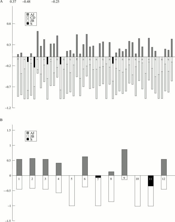
(A) Quantitative analysis from a series of patients with atrioventricular septal defect with common atrioventricular valvar orifices. The point zero corresponds to the level of atrioventricular junction. (B) Similar quantitative analysis from the series of patients with atrioventricular septal defect and separate atrioventricular orifices for right and left ventricles. AJ, dimension of the atrial component above the level of the junction; CD, ventricular component; JB, dimension of the atrial component below the plane of the junction; S, septum.
Figure 5 .

Apical four chamber view (A) from a patient with common atrioventricular junction and common valvar orifice. The plane of the atrioventricular junction is marked (< >). The lower edge of the atrial septum is below the level of the junction (arrow). (B) Similar cut from a patient with common atrioventricular junction, but separate right and left atrioventricular valvar orifices. As in (A), the atrial septum extends below (arrow) the level of the atrioventricular junction. AS, atrial septum; LA, left atrium; LV, left ventricle; RA, right atrium; RV, right ventricle; S, ventricular septum.
Selected References
These references are in PubMed. This may not be the complete list of references from this article.
- Anderson R. H., Ho S. Y., Falcao S., Daliento L., Rigby M. L. The diagnostic features of atrioventricular septal defect with common atrioventricular junction. Cardiol Young. 1998 Jan;8(1):33–49. doi: 10.1017/s1047951100004613. [DOI] [PubMed] [Google Scholar]
- Becker A. E., Anderson R. H. Atrioventricular septal defects: What's in a name? J Thorac Cardiovasc Surg. 1982 Mar;83(3):461–469. [PubMed] [Google Scholar]
- Cabrera A., Pastor E., Galdeano J. M., Modesto C., Cabrera J. A., Alcibar J., Peña R. Cross-sectional echocardiography in the diagnosis of atrioventricular septal defect. Int J Cardiol. 1990 Jul;28(1):19–23. doi: 10.1016/0167-5273(90)90004-o. [DOI] [PubMed] [Google Scholar]
- Cohen M. S., Jacobs M. L., Weinberg P. M., Rychik J. Morphometric analysis of unbalanced common atrioventricular canal using two-dimensional echocardiography. J Am Coll Cardiol. 1996 Oct;28(4):1017–1023. doi: 10.1016/s0735-1097(96)00262-8. [DOI] [PubMed] [Google Scholar]
- Ho S. Y., Russell G., Gerlis L. M. Atrioventricular septal defect with intact septal structures in a 74-year-old. Int J Cardiol. 1990 Mar;26(3):371–373. doi: 10.1016/0167-5273(90)90097-o. [DOI] [PubMed] [Google Scholar]
- Penkoske P. A., Neches W. H., Anderson R. H., Zuberbuhler J. R. Further observations on the morphology of atrioventricular septal defects. J Thorac Cardiovasc Surg. 1985 Oct;90(4):611–622. [PubMed] [Google Scholar]
- Sigfússon G., Ettedgui J. A., Silverman N. H., Anderson R. H. Is a cleft in the anterior leaflet of an otherwise normal mitral valve an atrioventricular canal malformation? J Am Coll Cardiol. 1995 Aug;26(2):508–515. doi: 10.1016/0735-1097(95)80030-k. [DOI] [PubMed] [Google Scholar]
- Silverman N. H., Ho S. Y., Anderson R. H., Smith A., Wilkinson J. L. Atrioventricular septal defect with intact atrial and ventricular septal structures. Int J Cardiol. 1984 May;5(5):567–573. doi: 10.1016/0167-5273(84)90167-0. [DOI] [PubMed] [Google Scholar]
- Smallhorn J. F., Tommasini G., Anderson R. H., Macartney F. J. Assessment of atrioventricular septal defects by two dimensional echocardiography. Br Heart J. 1982 Feb;47(2):109–121. doi: 10.1136/hrt.47.2.109. [DOI] [PMC free article] [PubMed] [Google Scholar]
- Smallhorn J. F., de Leval M., Stark J., Somerville J., Taylor J. F., Anderson R. H., Macartney F. J. Isolated anterior mitral cleft. Two dimensional echocardiographic assessment and differentiation from "clefts" associated with atrioventricular septal defect. Br Heart J. 1982 Aug;48(2):109–116. doi: 10.1136/hrt.48.2.109. [DOI] [PMC free article] [PubMed] [Google Scholar]