
Figure 3 .
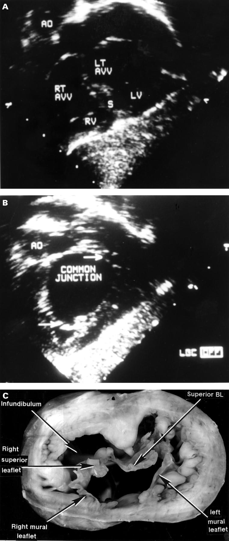
Comparable echocardiographic cuts (A, B) and a cross section from the heart of a different patient (C) showing the junctional arrangement in atrioventricular septal defect with common atrioventricular junction, but with separate valvar orifices for the right and left ventricles. The more distal echo cut (A) shows the common junction relative to the ventricular septum(s). As shown by the more proximal cut (B), at the level of the defect itself, the septum is not seen and the junction is obviously common (between arrows). This is confirmed by the cut of the postmortem specimen. AO, aorta; BL, bridging leaflet; LV, left ventricle; RT AVV, LI AVV, right and left atrioventricular valves; RV, right ventricle.