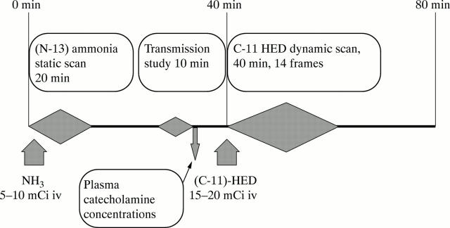
Figure 2 .
Imaging protocol. After positioning in the tomograph, a bolus injection of 185-370 MBq (5-10 mCi) of N-13 ammonia was followed by a 20 minute static acquisition, commencing three minutes after injection. Then a 10 minute transmission study was acquired. After waiting one hour for N-13 decay, neuronal imaging of the heart was performed after bolus injection of 740 MBq (20 mCi) of C-11 hydroxyephedrine, with a subsequent 40 minute dynamic data acquisition in frame mode (14 frames) to determine tracer activity in both blood and myocardium.

