Abstract
An electrophysiological study was performed in a 61 year old man with Wolff- Parkinson-White (WPW) syndrome. At baseline, neither ventricular nor supraventricular tachycardias could be induced. During isoprenaline infusion, ventricular tachycardia originating from the right ventricular outflow tract (RVOT) with a cycle length of 280 ms was induced and subsequently atrioventricular reentrant tachycardia (AVRT) with a cycle length of 300 ms using an accessory pathway in the left free wall appeared. During these tachycardias, AVRT was entrained by ventricular tachycardia. The earliest ventricular activation site during the ventricular tachycardia was determined to be the RVOT site and a radiofrequency current at 30 W successfully ablated the ventricular tachycardia at this site. The left free wall accessory pathway was also successfully ablated during right ventricular pacing. The coexistence of WPW syndrome and cathecolamine sensitive ventricular tachycardia originating from the RVOT has rarely been reported. Furthermore, the tachycardias were triggered by previous tachycardias. Keywords: double tachycardia; Wolff-Parkinson-White syndrome; radiofrequency catheter ablation; arrhythmias
Full Text
The Full Text of this article is available as a PDF (114.9 KB).
Figure 1 .
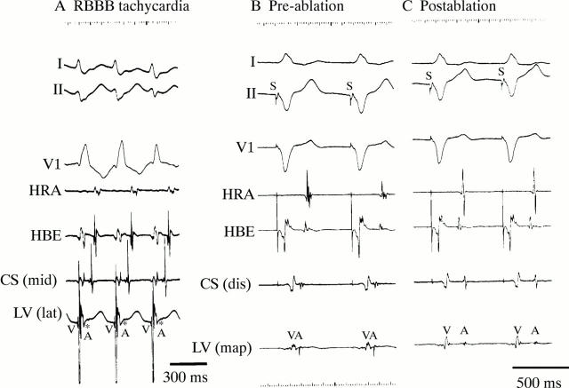
Intracardiac recordings during RBBB tachycardia and during ventricular pacing before and after ablation. Surface leads I, II, and V1 are shown with intracardiac electrograms from the high right atrium (HRA), the His bundle electrogram recording site (HBE), coronary sinus (CS), and the left ventricle (LV). (A) AVRT with RBBB morphology at a cycle length of 300 ms are seen. The earliest site of retrograde atrial conduction was observed at the left lateral site of the subvalvar portion of the mitral valve. The asterisks show the retrograde atrial activity. (B) and (C) The sequence of the ventriculoatrial conduction changed after the ablation. mid, middle portion; lat, lateral portion; dis, distal portion; map, mapping catheter.
Figure 2 .
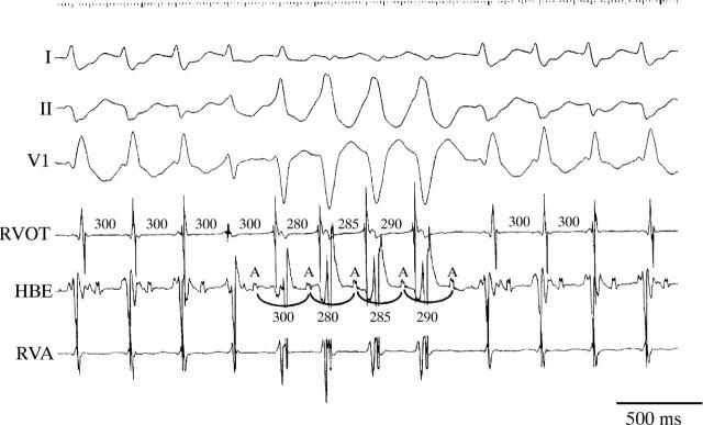
Intracardiac recording during double tachycardia. Surface leads I, II, and V1 are shown with intracardiac electrograms from the RVOT, the His bundle electrogram recording site (HBE), and the right ventricular apex (RVA). AVRT with RBBB morphology at a cycle length of 300 ms and non-sustained ventricular tachycardia with LBBB morphology at a cycle length of 280-290 ms are seen. Transient entrainment phenomenon of the AVRT by non-sustained ventricular tachycardia is observed.
Figure 3 .
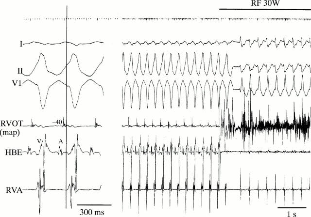
Radiofrequency catheter ablation (RF) of the origin of ventricular tachycardia in the right ventricular outflow tract. (Left) The earliest activation site was determined. The local electrogram at the site preceded the onset of the QRS complex by 40 ms. (Right) RF current at 30 W was delivered to the earliest activation site, the ventricular tachycardia was successfully ablated and the AVRT appeared. map, mapping catheter; HBE, His bundle electrogram; RVA, right ventricular apex.


