Abstract
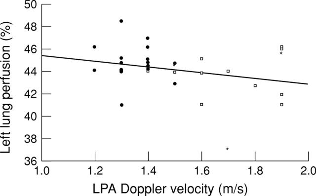
OBJECTIVE—To evaluate relative lung perfusion following complete occlusion of persistent arterial duct with detachable Cook coils. METHODS—Ductal occlusion using detachable coils was performed in 35 patients (median age 3.9 years, range 0.5 to 16; 32 native ducts, three patients with previous devices). If the duct could be crossed with a 0.035 inch guidewire and a 4 F catheter after coil implantation, a further coil was implanted. Between one and seven coils were used (median two). RESULTS—Complete ductal occlusion was confirmed by echocardiography 24 hours after the procedure in all patients. Lung perfusion scans were performed three months after the procedure in 33 of 35 patients (two older patients with a single coil each did not attend). Decreased perfusion to the left lung (defined as < 40% of total lung flow) was observed in only one patient, who had previously had a 17 mm Rashkind umbrella implanted. There was no correlation between left lung perfusion and peak left pulmonary artery Doppler velocities (r = 0.27 and p = 0.125 for the entire group; r = 0.29 and p = 0.124 after excluding patients with previous devices). CONCLUSIONS—Coil occlusion is effective in achieving complete closure of the duct. An aggressive approach using multiple coils did not compromise perfusion to the left lung. Keywords: arterial duct; coil occlusion; lung perfusion
Full Text
The Full Text of this article is available as a PDF (109.1 KB).
Figure 1 .
Scatter diagram of left lung perfusion as percentage of total lung flow (y axis) and peak left pulmonary artery (LPA) Doppler velocity in m/s (x axis). Filled symbols denote patients without coil protrusion into the left pulmonary artery on cross sectional echocardio-graphy, empty symbols denote patients with coil protrusion. Asterisks denote patients with previous devices implanted in the duct. The line represents the least squares regression line (r = 0.27; p = 0.125). Formula: y = −2.5 + 47.8 (95% confidence interval of coefficient: −5.7 to 0.73; 95% confidence interval of constant: 43.0 to 52.6).
Selected References
These references are in PubMed. This may not be the complete list of references from this article.
- Dessy H., Hermus J. P., van den Heuvel F., Oei H. Y., Krenning E. P., Hess J. Echocardiographic and radionuclide pulmonary blood flow patterns after transcatheter closure of patent ductus arteriosus. Circulation. 1996 Jul 15;94(2):126–129. doi: 10.1161/01.cir.94.2.126. [DOI] [PubMed] [Google Scholar]
- Evangelista J. K., Hijazi Z. M., Geggel R. L., Oates E., Fulton D. R. Effect of multiple coil closure of patent ductus arteriosus on blood flow to the left lung as determined by lung perfusion scans. Am J Cardiol. 1997 Jul 15;80(2):242–244. doi: 10.1016/s0002-9149(97)00335-4. [DOI] [PubMed] [Google Scholar]
- Fadley F., al-Halees Z., Galal O., Kumar N., Wilson N. Left pulmonary artery stenosis after transcatheter occlusion of persistent arterial duct. Lancet. 1993 Feb 27;341(8844):559–560. doi: 10.1016/0140-6736(93)90321-7. [DOI] [PubMed] [Google Scholar]
- Hijazi Z. M., Geggel R. L. Results of anterograde transcatheter closure of patent ductus arteriosus using single or multiple Gianturco coils. Am J Cardiol. 1994 Nov 1;74(9):925–929. doi: 10.1016/0002-9149(94)90588-6. [DOI] [PubMed] [Google Scholar]
- Hijazi Z. M., Geggel R. L. Transcatheter closure of large patent ductus arteriosus (> or = 4 mm) with multiple Gianturco coils: immediate and mid-term results. Heart. 1996 Dec;76(6):536–540. doi: 10.1136/hrt.76.6.536. [DOI] [PMC free article] [PubMed] [Google Scholar]
- Hosking M. C., Benson L. N., Musewe N., Dyck J. D., Freedom R. M. Transcatheter occlusion of the persistently patent ductus arteriosus. Forty-month follow-up and prevalence of residual shunting. Circulation. 1991 Dec;84(6):2313–2317. doi: 10.1161/01.cir.84.6.2313. [DOI] [PubMed] [Google Scholar]
- Krichenko A., Benson L. N., Burrows P., Möes C. A., McLaughlin P., Freedom R. M. Angiographic classification of the isolated, persistently patent ductus arteriosus and implications for percutaneous catheter occlusion. Am J Cardiol. 1989 Apr 1;63(12):877–880. doi: 10.1016/0002-9149(89)90064-7. [DOI] [PubMed] [Google Scholar]
- Lloyd T. R., Fedderly R., Mendelsohn A. M., Sandhu S. K., Beekman R. H., 3rd Transcatheter occlusion of patent ductus arteriosus with Gianturco coils. Circulation. 1993 Oct;88(4 Pt 1):1412–1420. doi: 10.1161/01.cir.88.4.1412. [DOI] [PubMed] [Google Scholar]
- Nykanen D. G., Hayes A. M., Benson L. N., Freedom R. M. Transcatheter patent ductus arteriosus occlusion: application in the small child. J Am Coll Cardiol. 1994 Jun;23(7):1666–1670. doi: 10.1016/0735-1097(94)90672-6. [DOI] [PubMed] [Google Scholar]
- Perry S. B., Lock J. E. Front-loading of double-umbrella devices, a new technique for umbrella delivery for closing cardiovascular defects. Am J Cardiol. 1992 Oct 1;70(9):917–920. doi: 10.1016/0002-9149(92)90738-k. [DOI] [PubMed] [Google Scholar]
- Shim D., Fedderly R. T., Beekman R. H., 3rd, Ludomirsky A., Young M. L., Schork A., Lloyd T. R. Follow-up of coil occlusion of patent ductus arteriosus. J Am Coll Cardiol. 1996 Jul;28(1):207–211. doi: 10.1016/0735-1097(96)00107-6. [DOI] [PubMed] [Google Scholar]
- Tometzki A. J., Arnold R., Peart I., Sreeram N., Abdulhamed J. M., Godman M. J., Patel R. G., Kitchiner D. J., Bu'Lock F. A., Walsh K. P. Transcatheter occlusion of the patent ductus arteriosus with Cook detachable coils. Heart. 1996 Dec;76(6):531–535. doi: 10.1136/hrt.76.6.531. [DOI] [PMC free article] [PubMed] [Google Scholar]


