Abstract
OBJECTIVE—To determine whether restriction at the atrial septum in the newborn with hypoplastic left heart syndrome can be predicted accurately by examining the pattern of pulmonary venous flow in the fetus. A restrictive atrial septum can contribute to haemodynamic instability before surgery for this lesion and has been associated with an increased mortality. DESIGN—Pulmonary venous pulsed Doppler tracings were compared between fetuses with hypoplastic left heart syndrome and controls. The size of the atrial septal defect on the postnatal echocardiogram was graded according to the degree of restriction. Pulsed Doppler tracings of pulmonary venous blood flow were obtained in 18 fetuses with left atrial outflow atresia and compared with 77 controls, adjusted for gestational age. Postnatal echocardiograms were available for analysis in 13 of 18 neonates. SETTING—A tertiary referral centre for fetal cardiology and paediatric cardiac surgery. RESULTS—Fetuses with hypoplastic left heart syndrome were different from controls in all pulmonary vein indices measured. As assessed from the postnatal echocardiogram, there were seven fetuses with a restrictive atrial septum. In these fetuses, the systolic flow velocity (p < 0.01), S/D ratio (p < 0.01), and peak reversal wave (p < 0.001) in the pulmonary vein tracing showed a good correlation with the degree of restriction. CONCLUSIONS—The Doppler pattern of pulmonary venous flow in the fetus with hypoplastic left heart syndrome appears to be a reliable predictor of restriction of the atrial septum in the neonate. This may help in the immediate post-delivery management of these infants before surgery. Keywords: fetus; congenital heart defects; echocardiography; risk factors
Full Text
The Full Text of this article is available as a PDF (118.8 KB).
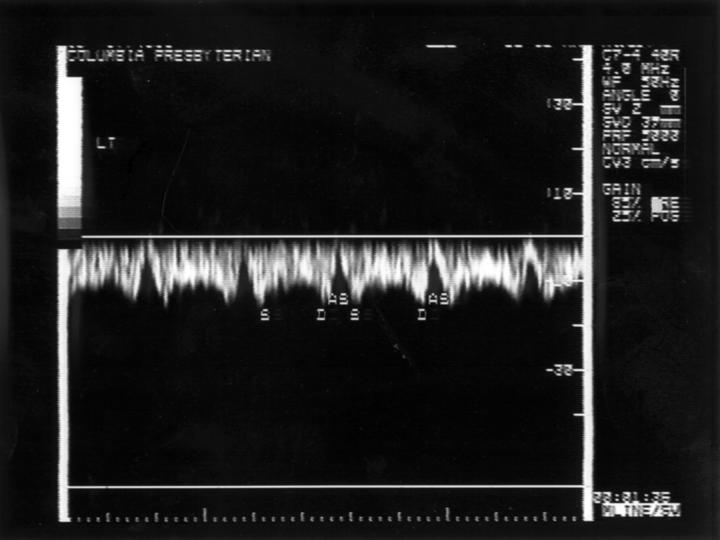
Figure 1 .
The normal pattern of pulmonary vein flow in a fetus. There is a systolic peak (S) followed by an early diastolic peak (D) and flow cessation or reversal during late diastole or atrial systole (AS). The systolic and diastolic velocities are similar and there is a mild rise in velocity of forward flow with advancing gestation with a range of 10 cm/s to 40 cm/s between 16 weeks and term.
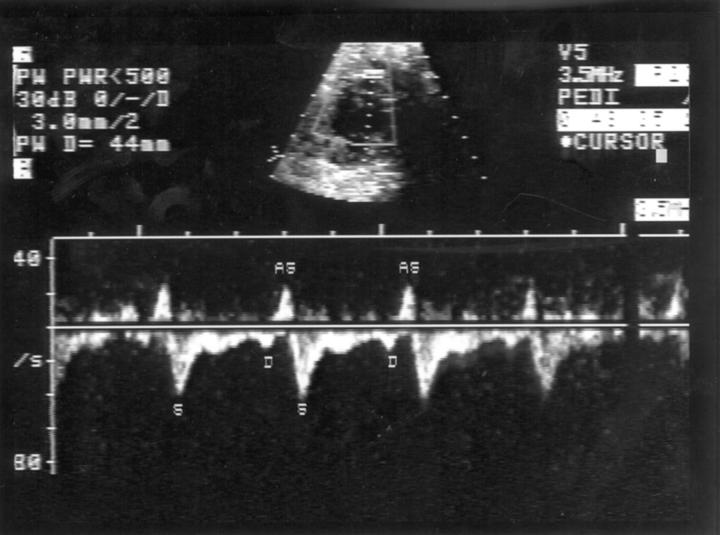
Figure 2 .
The pattern of pulmonary venous flow in a fetus with the hypoplastic left heart syndrome who had a severely restrictive atrial septum postnatally. The systolic and reverse waves were higher than normal and the diastolic lower than normal for gestational age. The systolic/diastolic ratio was also abnormal.
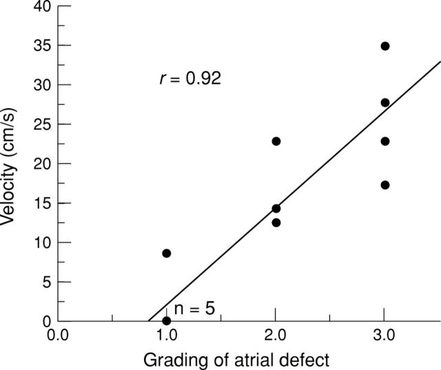
Figure 3 .
The velocity of the peak reversal wave plotted against the grading of the degree of restriction at the atrial septum on the postnatal echocardiogram, with grade 1 being unrestrictive and grade 3 the most restrictive. Note that there were five fetuses with no flow reversal, represented by a single point on the baseline.
Selected References
These references are in PubMed. This may not be the complete list of references from this article.
- Allan L. D., Sharland G. K., Milburn A., Lockhart S. M., Groves A. M., Anderson R. H., Cook A. C., Fagg N. L. Prospective diagnosis of 1,006 consecutive cases of congenital heart disease in the fetus. J Am Coll Cardiol. 1994 May;23(6):1452–1458. doi: 10.1016/0735-1097(94)90391-3. [DOI] [PubMed] [Google Scholar]
- Basnight M. A., Gonzalez M. S., Kershenovich S. C., Appleton C. P. Pulmonary venous flow velocity: relation to hemodynamics, mitral flow velocity and left atrial volume, and ejection fraction. J Am Soc Echocardiogr. 1991 Nov-Dec;4(6):547–558. doi: 10.1016/s0894-7317(14)80213-7. [DOI] [PubMed] [Google Scholar]
- Better D. J., Kaufman S., Allan L. D. The normal pattern of pulmonary venous flow on pulsed Doppler examination of the human fetus. J Am Soc Echocardiogr. 1996 May-Jun;9(3):281–285. doi: 10.1016/s0894-7317(96)90141-8. [DOI] [PubMed] [Google Scholar]
- Bove E. L., Lloyd T. R. Staged reconstruction for hypoplastic left heart syndrome. Contemporary results. Ann Surg. 1996 Sep;224(3):387–395. doi: 10.1097/00000658-199609000-00015. [DOI] [PMC free article] [PubMed] [Google Scholar]
- Chang A. C., Huhta J. C., Yoon G. Y., Wood D. C., Tulzer G., Cohen A., Mennuti M., Norwood W. I. Diagnosis, transport, and outcome in fetuses with left ventricular outflow tract obstruction. J Thorac Cardiovasc Surg. 1991 Dec;102(6):841–848. [PubMed] [Google Scholar]
- Keren G., Sherez J., Megidish R., Levitt B., Laniado S. Pulmonary venous flow pattern--its relationship to cardiac dynamics. A pulsed Doppler echocardiographic study. Circulation. 1985 Jun;71(6):1105–1112. doi: 10.1161/01.cir.71.6.1105. [DOI] [PubMed] [Google Scholar]
- Klein A. L., Tajik A. J. Doppler assessment of pulmonary venous flow in healthy subjects and in patients with heart disease. J Am Soc Echocardiogr. 1991 Jul-Aug;4(4):379–392. doi: 10.1016/s0894-7317(14)80448-3. [DOI] [PubMed] [Google Scholar]
- Laudy J. A., Huisman T. W., de Ridder M. A., Wladimiroff J. W. Normal fetal pulmonary venous blood flow velocity. Ultrasound Obstet Gynecol. 1995 Oct;6(4):277–281. doi: 10.1046/j.1469-0705.1995.06040277.x. [DOI] [PubMed] [Google Scholar]
- Montaña E., Khoury M. J., Cragan J. D., Sharma S., Dhar P., Fyfe D. Trends and outcomes after prenatal diagnosis of congenital cardiac malformations by fetal echocardiography in a well defined birth population, Atlanta, Georgia, 1990-1994. J Am Coll Cardiol. 1996 Dec;28(7):1805–1809. doi: 10.1016/S0735-1097(96)00381-6. [DOI] [PubMed] [Google Scholar]
- Rasanen J., Wood D. C., Weiner S., Ludomirski A., Huhta J. C. Role of the pulmonary circulation in the distribution of human fetal cardiac output during the second half of pregnancy. Circulation. 1996 Sep 1;94(5):1068–1073. doi: 10.1161/01.cir.94.5.1068. [DOI] [PubMed] [Google Scholar]
- Stavri G. T., Zachary I. C., Baskerville P. A., Martin J. F., Erusalimsky J. D. Basic fibroblast growth factor upregulates the expression of vascular endothelial growth factor in vascular smooth muscle cells. Synergistic interaction with hypoxia. Circulation. 1995 Jul 1;92(1):11–14. doi: 10.1161/01.cir.92.1.11. [DOI] [PubMed] [Google Scholar]
- Sutton M. S., Groves A., MacNeill A., Sharland G., Allan L. Assessment of changes in blood flow through the lungs and foramen ovale in the normal human fetus with gestational age: a prospective Doppler echocardiographic study. Br Heart J. 1994 Mar;71(3):232–237. doi: 10.1136/hrt.71.3.232. [DOI] [PMC free article] [PubMed] [Google Scholar]


