Abstract
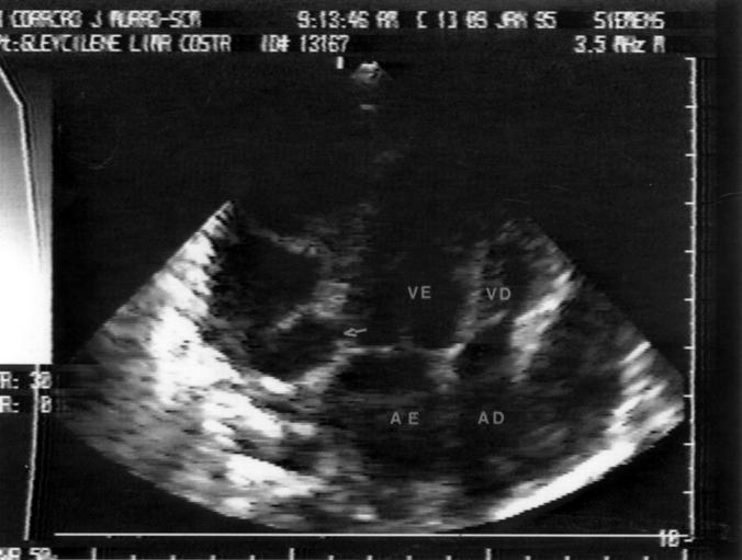
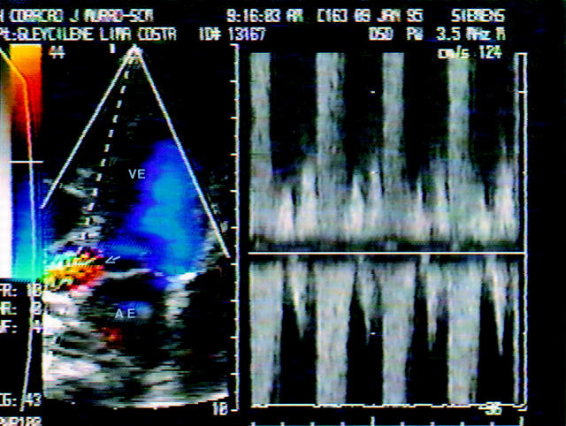
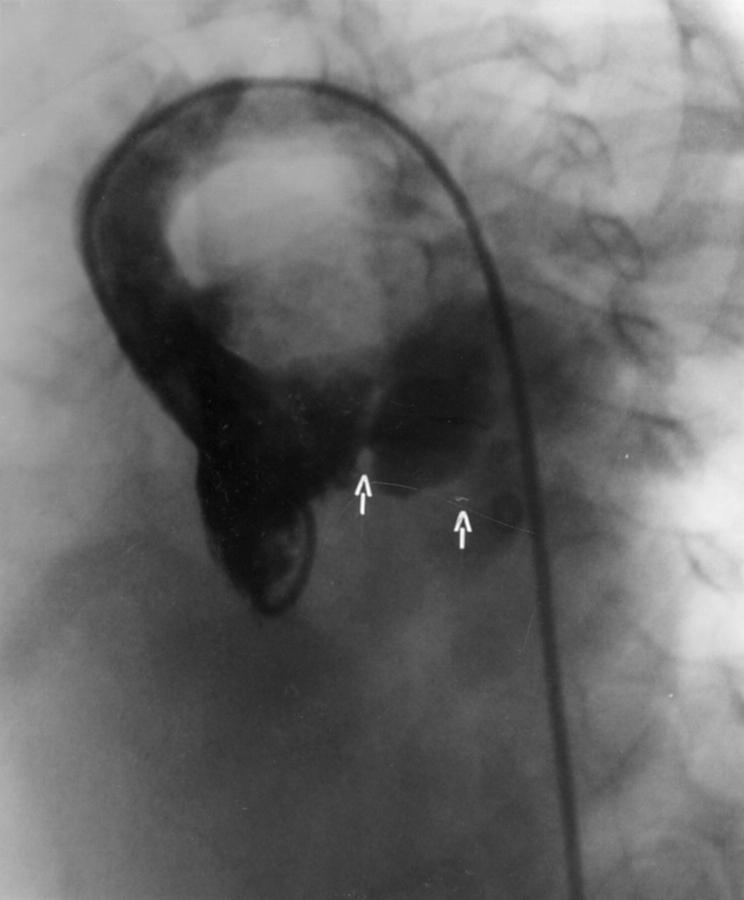
Cross sectional echocardiography demonstrated a pseudoaneurysm of the left ventricular posterolateral wall close to the atrioventricular junction in a 4 year old girl with infective pericarditis complicating lobar pneumonia. Colour flow Doppler demonstrated bidirectional flow across the communication hole. Surgical resection was successful. Keywords: pseudoaneurysm; pericarditis; echocardiography
Full Text
The Full Text of this article is available as a PDF (91.2 KB).
Figure 1 .
Cross sectional echocardiography (apical four chamber view) showing large pericardial effusion and the communication between the left ventricle and the pseudoaneurysm (arrow). VE, left ventricle; VD, right ventricle; AE, left atrium; AD, right atrium.
Figure 2 .
Colour flow mapping and pulsed Doppler recording at the origin of the pseudoaneurysm (arrow). VE, left ventricle; AE, left atrium.
Figure 3 .
Left ventriculogram in the left anterior oblique view showing a large aneurysm of the posterolateral wall (arrows).


