Abstract
OBJECTIVE—To analyse the prodrome of acute myocardial infarction in relation to the plaque morphology underlying the infarct. DESIGN—A retrospective investigation of the relation between rupture and erosion of coronary atheromatous plaques and the clinical characteristics of acute myocardial infarction. The coronary arteries of 100 patients who died from acute myocardial infarction were cut transversely at 3 mm intervals. Segments with a stenosis were examined microscopically at 5 µm intervals. The clinical features of the infarction were obtained from the medical records. RESULTS—A deep intimal rupture was encountered in 81 plaques, whereas 19 had superficial erosions only. There were no differences in the location of infarction, the incidence of hypertension, diabetes mellitus, or hyperlipidaemia, diameter stenosis of the infarcted related artery, Killip class, Forrester's haemodynamic subset, or peak creatine kinase between plaque rupture and plaque erosion groups. The presence of plaque rupture was associated with significantly greater incidences of leucocytosis, current smoking, and sudden or unstable onset of acute coronary syndrome. In patients with unstable preinfarction angina, new onset rest angina rather than worsening angina tended to develop more often in the plaque rupture group than in the plaque erosion group (p = 0.08). CONCLUSIONS—Plaque rupture causes the sudden onset of acute myocardial infarction or unstable preinfarction angina, which may be aggravated by smoking and inflammation. Keywords: acute coronary syndrome; plaque rupture; white blood cell; preinfarction angina
Full Text
The Full Text of this article is available as a PDF (102.3 KB).
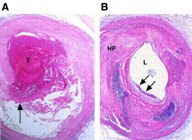
Figure 1 .
(A) A coronary plaque rupture was found in a patient who died one week after the onset of acute myocardial infarction. At lower magnification the rupture site (arrow) is seen at the shoulder lesion, which is considered to have had a thin fibrous cap. An occlusive thrombus (T) was present (haematoxylin and eosin stain). (B) A coronary plaque erosion was found in a patient who died two weeks after the onset of acute myocardial infarction. At lower magnification, a concentric hard plaque (HP) which includes focal calcification was present. The luminal (L) plaque surface was in contact with non-occlusive thrombus (arrows) (haematoxylin and eosin stain).
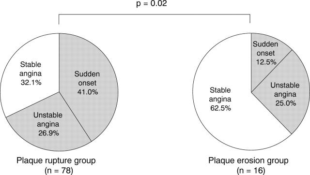
Figure 2 .
Pie charts showing the status of preinfarction angina. The patients with plaque rupture had a significantly higher incidence of sudden onset and unstable angina than those with plaque erosion.
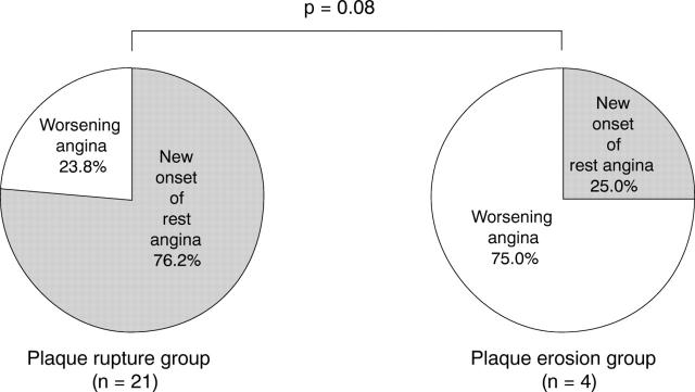
Figure 3 .
Pie charts showing the status of unstable angina. The patients with plaque rupture had a higher incidence of new onset of rest angina than those with plaque erosion, but the difference was not significant.
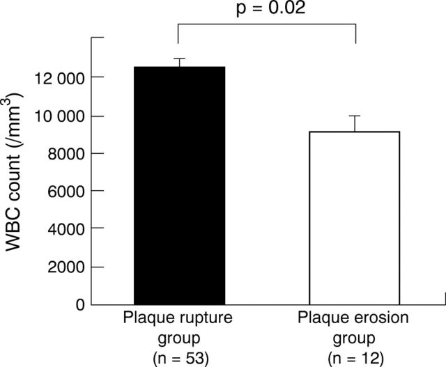
Figure 4 .
White blood cell count within six hours of the onset of acute myocardial infarction. Error bars = SEM.
Selected References
These references are in PubMed. This may not be the complete list of references from this article.
- Annex B. H., Denning S. M., Channon K. M., Sketch M. H., Jr, Stack R. S., Morrissey J. H., Peters K. G. Differential expression of tissue factor protein in directional atherectomy specimens from patients with stable and unstable coronary syndromes. Circulation. 1995 Feb 1;91(3):619–622. doi: 10.1161/01.cir.91.3.619. [DOI] [PubMed] [Google Scholar]
- Burke A. P., Farb A., Malcom G. T., Liang Y. H., Smialek J., Virmani R. Coronary risk factors and plaque morphology in men with coronary disease who died suddenly. N Engl J Med. 1997 May 1;336(18):1276–1282. doi: 10.1056/NEJM199705013361802. [DOI] [PubMed] [Google Scholar]
- Burke A. P., Farb A., Malcom G. T., Liang Y., Smialek J., Virmani R. Effect of risk factors on the mechanism of acute thrombosis and sudden coronary death in women. Circulation. 1998 Jun 2;97(21):2110–2116. doi: 10.1161/01.cir.97.21.2110. [DOI] [PubMed] [Google Scholar]
- Capone G., Wolf N. M., Meyer B., Meister S. G. Frequency of intracoronary filling defects by angiography in angina pectoris at rest. Am J Cardiol. 1985 Sep 1;56(7):403–406. doi: 10.1016/0002-9149(85)90875-6. [DOI] [PubMed] [Google Scholar]
- Davies M. J., Thomas A. Thrombosis and acute coronary-artery lesions in sudden cardiac ischemic death. N Engl J Med. 1984 May 3;310(18):1137–1140. doi: 10.1056/NEJM198405033101801. [DOI] [PubMed] [Google Scholar]
- Falk E. Plaque rupture with severe pre-existing stenosis precipitating coronary thrombosis. Characteristics of coronary atherosclerotic plaques underlying fatal occlusive thrombi. Br Heart J. 1983 Aug;50(2):127–134. doi: 10.1136/hrt.50.2.127. [DOI] [PMC free article] [PubMed] [Google Scholar]
- Falk E., Shah P. K., Fuster V. Coronary plaque disruption. Circulation. 1995 Aug 1;92(3):657–671. doi: 10.1161/01.cir.92.3.657. [DOI] [PubMed] [Google Scholar]
- Farb A., Burke A. P., Tang A. L., Liang T. Y., Mannan P., Smialek J., Virmani R. Coronary plaque erosion without rupture into a lipid core. A frequent cause of coronary thrombosis in sudden coronary death. Circulation. 1996 Apr 1;93(7):1354–1363. doi: 10.1161/01.cir.93.7.1354. [DOI] [PubMed] [Google Scholar]
- Fuster V. Lewis A. Conner Memorial Lecture. Mechanisms leading to myocardial infarction: insights from studies of vascular biology. Circulation. 1994 Oct;90(4):2126–2146. doi: 10.1161/01.cir.90.4.2126. [DOI] [PubMed] [Google Scholar]
- Galis Z. S., Sukhova G. K., Lark M. W., Libby P. Increased expression of matrix metalloproteinases and matrix degrading activity in vulnerable regions of human atherosclerotic plaques. J Clin Invest. 1994 Dec;94(6):2493–2503. doi: 10.1172/JCI117619. [DOI] [PMC free article] [PubMed] [Google Scholar]
- Gertz S. D., Roberts W. C. Hemodynamic shear force in rupture of coronary arterial atherosclerotic plaques. Am J Cardiol. 1990 Dec 1;66(19):1368–1372. doi: 10.1016/0002-9149(90)91170-b. [DOI] [PubMed] [Google Scholar]
- Haverkate F., Thompson S. G., Pyke S. D., Gallimore J. R., Pepys M. B. Production of C-reactive protein and risk of coronary events in stable and unstable angina. European Concerted Action on Thrombosis and Disabilities Angina Pectoris Study Group. Lancet. 1997 Feb 15;349(9050):462–466. doi: 10.1016/s0140-6736(96)07591-5. [DOI] [PubMed] [Google Scholar]
- Horie T., Sekiguchi M., Hirosawa K. Coronary thrombosis in pathogenesis of acute myocardial infarction. Histopathological study of coronary arteries in 108 necropsied cases using serial section. Br Heart J. 1978 Feb;40(2):153–161. doi: 10.1136/hrt.40.2.153. [DOI] [PMC free article] [PubMed] [Google Scholar]
- Kovanen P. T., Kaartinen M., Paavonen T. Infiltrates of activated mast cells at the site of coronary atheromatous erosion or rupture in myocardial infarction. Circulation. 1995 Sep 1;92(5):1084–1088. doi: 10.1161/01.cir.92.5.1084. [DOI] [PubMed] [Google Scholar]
- Liuzzo G., Biasucci L. M., Rebuzzi A. G., Gallimore J. R., Caligiuri G., Lanza G. A., Quaranta G., Monaco C., Pepys M. B., Maseri A. Plasma protein acute-phase response in unstable angina is not induced by ischemic injury. Circulation. 1996 Nov 15;94(10):2373–2380. doi: 10.1161/01.cir.94.10.2373. [DOI] [PubMed] [Google Scholar]
- Moreno P. R., Bernardi V. H., López-Cuéllar J., Murcia A. M., Palacios I. F., Gold H. K., Mehran R., Sharma S. K., Nemerson Y., Fuster V. Macrophages, smooth muscle cells, and tissue factor in unstable angina. Implications for cell-mediated thrombogenicity in acute coronary syndromes. Circulation. 1996 Dec 15;94(12):3090–3097. doi: 10.1161/01.cir.94.12.3090. [DOI] [PubMed] [Google Scholar]
- Muller J. E., Abela G. S., Nesto R. W., Tofler G. H. Triggers, acute risk factors and vulnerable plaques: the lexicon of a new frontier. J Am Coll Cardiol. 1994 Mar 1;23(3):809–813. doi: 10.1016/0735-1097(94)90772-2. [DOI] [PubMed] [Google Scholar]
- Weiss S. J. Tissue destruction by neutrophils. N Engl J Med. 1989 Feb 9;320(6):365–376. doi: 10.1056/NEJM198902093200606. [DOI] [PubMed] [Google Scholar]
- Yutani C., Ishibashi-Ueda H., Konishi M., Shibata J., Arita M. Histopathological study of acute myocardial infarction and pathoetiology of coronary thrombosis: a comparative study in four districts in Japan. Jpn Circ J. 1987 Mar;51(3):352–361. doi: 10.1253/jcj.51.352. [DOI] [PubMed] [Google Scholar]
- van der Wal A. C., Becker A. E., van der Loos C. M., Das P. K. Site of intimal rupture or erosion of thrombosed coronary atherosclerotic plaques is characterized by an inflammatory process irrespective of the dominant plaque morphology. Circulation. 1994 Jan;89(1):36–44. doi: 10.1161/01.cir.89.1.36. [DOI] [PubMed] [Google Scholar]


