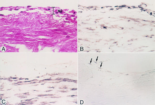
Figure 3 .
Micrographs of a vein graft at an anastomotic site, nine days after grafting (patient 5). Panels A-D represent serial sections. (A) Haematoxylin and eosin stain. Endothelial cells have been denuded. Early neointimal tissue (NI) is seen at the luminal surface of the graft. (B) Vimentin stain. Both spindle shaped cells and round cells in the neointima are positive. (C) HHF-35. The cells in the neointima do not stain. (D) PCNA. Some of the HHF-35 negative spindle shaped cells (arrows) stain positive. Original magnification × 580.

