

Full Text
The Full Text of this article is available as a PDF (243.5 KB).
Figure 1 .
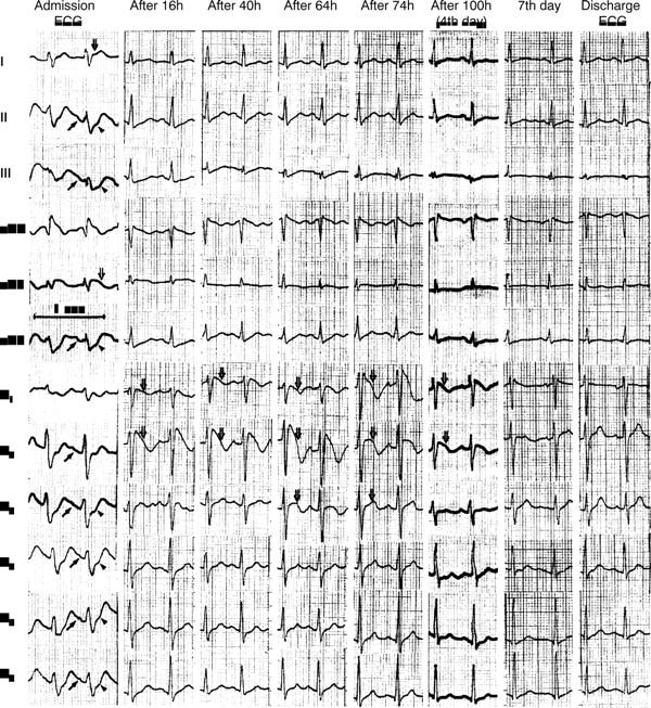
Serial ECGs recorded during hospitalisation. Admission ECG shows abnormal atrial and ventricular repolarisation. PR segment is depressed in II, III, aVF, V2 to V6 (small arrows), whereas ST segment elevation is evident in I and aVL (large arrows) with reciprocal ST depression in II, III, aVF, and V3 to V6 (arrowheads). The initial alterations do not exist in the second ECG, but new ST elevation with T wave inversion is obvious in V1 to V3 (large arrows). These become more prominent over time and persist for four days.