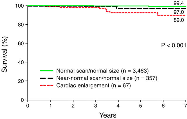
Figure 2: .
Cardiac survival in patients with intermediate risk exercise ECGs, subgrouped on the basis of their findings on perfusion imaging. The three subgroups shown—patients with normal perfusion scans and normal heart size; patients with near normal scans and normal heart size; and patients with cardiac enlargement—were significantly different from one another (p < 0.001). Both of the subgroups with normal heart size had a low risk of subsequent cardiac death, with an annual cardiac mortality of less than 0.5%. (Reproduced from Gibbons et al 12 with permission of the American Heart Association.)

