Full Text
The Full Text of this article is available as a PDF (223.3 KB).
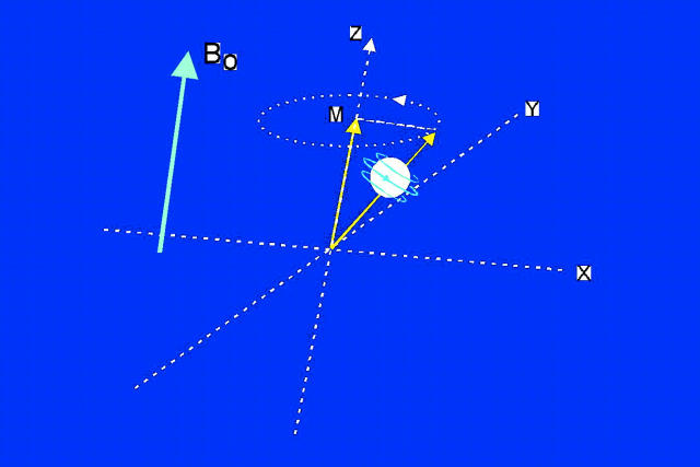
Figure 1: .
Spin angular moment causing a magnetic dipole. Bo, external magnetic field which causes spin to precess at an angle to Bo. M, net tissue magnetisation aligned along Z axis.
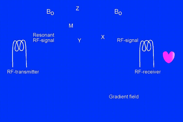
Figure 2: .
The patient (heart) is placed within a strong external magnetic field (Bo). The RF transmitter rotates the net tissue magnetisation in the transverse plane, and after termination, relaxation occurs which emits a signal detected by the RF receiver. The gradient coils produce a supplemental magnetic field gradient to allow precise location of the excited protons. The received signals have certain signal intensity (brightness) and location, both of which are processed to form the desired image.
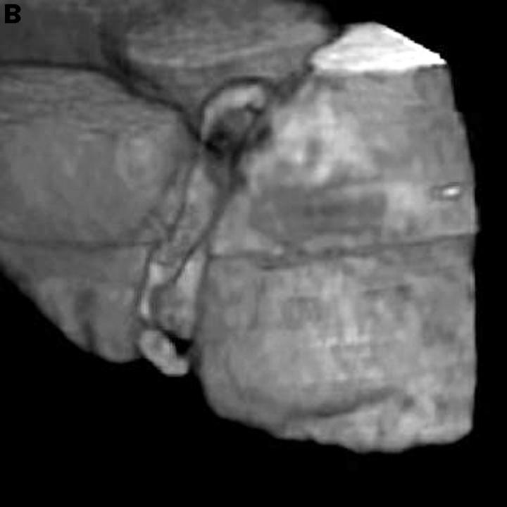
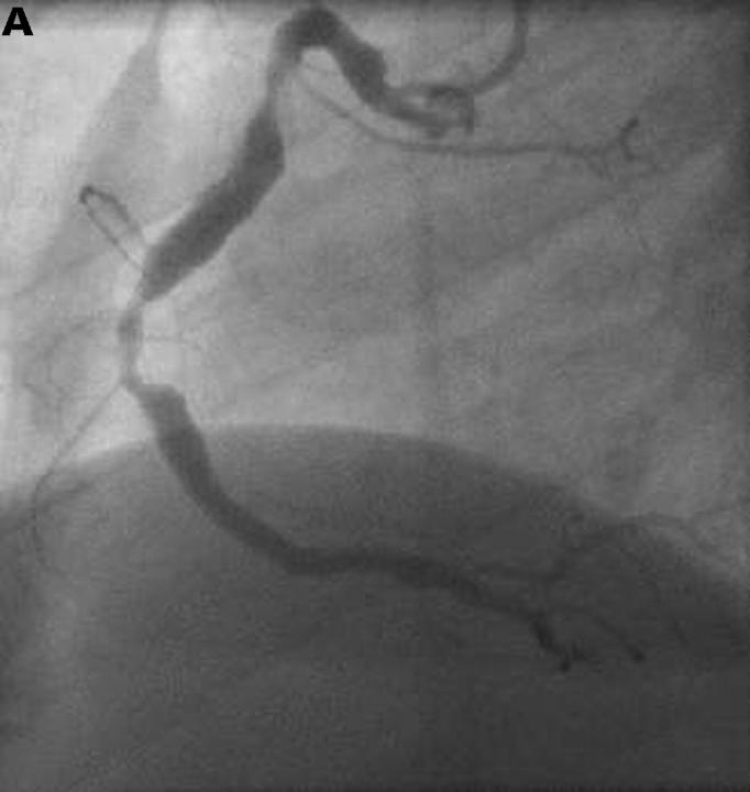
Figure 3: .
Visualisation of the right coronary artery with (A) a fast sequence MR coronary angiography technique, and (B) three dimensional reconstruction. Two significant stenoses are clearly visible.
Figure 4: .
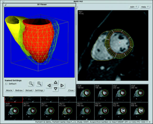
Computer display of the MASS analytical software package. The small images along the bottom represent the individual frames over a cardiac cycle at a certain anatomical level. The left ventricular endocardial and epicardial contours were generated using semiautomatic contour detection. From the contours in all the frames and slices, a three dimensional model can be reconstructed that can also be used as a functional display representing regional wall thickening/thinning. Reproduced from van der Geest et al. J Comput Assist Tomogr 1997;21:756-65, with permission of the publishers.
Figure 5: .
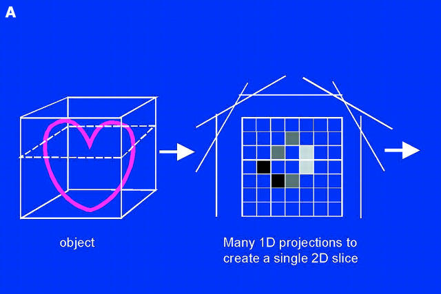
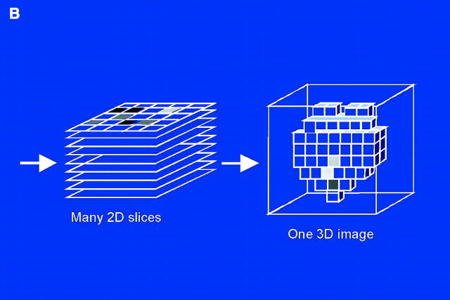
Schematic of EBT scanning to reconstruct a three dimensional image. ID, one dimensional; 2D, two dimensional; 3D, 3 dimensional.
Figure 6: .
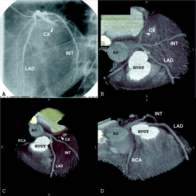
EBT of coronary arteries with view from top (B), from a more anterior angle (C), and from a lateral angle (D). The left circumflex (CX) artery is totally occluded. A: corresponding coronary angiography. AO, aorta; INT, intermediate coronary branch; LAD, left anterior descending coronary artery; RCA, right coronary artery, RVOT, right ventricular outflow tract.
Figure 7: .
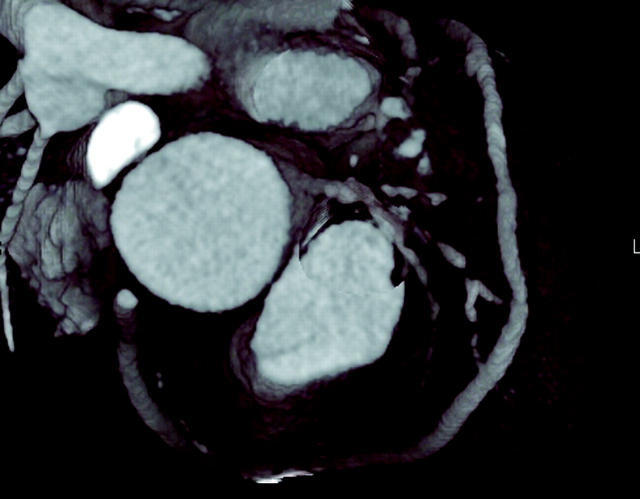
Visualisation of sequential venous bypass graft without significant stenoses.
Figure 8: .
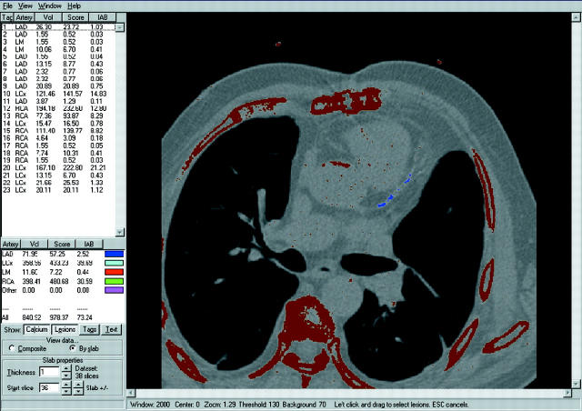
Example of coronary calcification score. Blue dots in the left anterior descending artery represent calcification.
Selected References
These references are in PubMed. This may not be the complete list of references from this article.
- Agatston A. S., Janowitz W. R., Hildner F. J., Zusmer N. R., Viamonte M., Jr, Detrano R. Quantification of coronary artery calcium using ultrafast computed tomography. J Am Coll Cardiol. 1990 Mar 15;15(4):827–832. doi: 10.1016/0735-1097(90)90282-t. [DOI] [PubMed] [Google Scholar]
- Baer F. M., Theissen P., Schneider C. A., Voth E., Sechtem U., Schicha H., Erdmann E. Dobutamine magnetic resonance imaging predicts contractile recovery of chronically dysfunctional myocardium after successful revascularization. J Am Coll Cardiol. 1998 Apr;31(5):1040–1048. doi: 10.1016/s0735-1097(98)00032-1. [DOI] [PubMed] [Google Scholar]
- Brown M. A., Semelka R. C. MR imaging abbreviations, definitions, and descriptions: a review. Radiology. 1999 Dec;213(3):647–662. doi: 10.1148/radiology.213.3.r99dc18647. [DOI] [PubMed] [Google Scholar]
- Geskin G., Kramer C. M., Rogers W. J., Theobald T. M., Pakstis D., Hu Y. L., Reichek N. Quantitative assessment of myocardial viability after infarction by dobutamine magnetic resonance tagging. Circulation. 1998 Jul 21;98(3):217–223. doi: 10.1161/01.cir.98.3.217. [DOI] [PubMed] [Google Scholar]
- Hoffmann U., Globits S., Frank H. Cardiac and paracardiac masses. Current opinion on diagnostic evaluation by magnetic resonance imaging. Eur Heart J. 1998 Apr;19(4):553–563. doi: 10.1053/euhj.1997.0788. [DOI] [PubMed] [Google Scholar]
- Hundley W. G., Hamilton C. A., Thomas M. S., Herrington D. M., Salido T. B., Kitzman D. W., Little W. C., Link K. M. Utility of fast cine magnetic resonance imaging and display for the detection of myocardial ischemia in patients not well suited for second harmonic stress echocardiography. Circulation. 1999 Oct 19;100(16):1697–1702. doi: 10.1161/01.cir.100.16.1697. [DOI] [PubMed] [Google Scholar]
- Manning W. J., Li W., Edelman R. R. A preliminary report comparing magnetic resonance coronary angiography with conventional angiography. N Engl J Med. 1993 Mar 25;328(12):828–832. doi: 10.1056/NEJM199303253281202. [DOI] [PubMed] [Google Scholar]
- Moshage W. E., Achenbach S., Seese B., Bachmann K., Kirchgeorg M. Coronary artery stenoses: three-dimensional imaging with electrocardiographically triggered, contrast agent-enhanced, electron-beam CT. Radiology. 1995 Sep;196(3):707–714. doi: 10.1148/radiology.196.3.7644633. [DOI] [PubMed] [Google Scholar]
- Post J. C., van Rossum A. C., Bronzwaer J. G., de Cock C. C., Hofman M. B., Valk J., Visser C. A. Magnetic resonance angiography of anomalous coronary arteries. A new gold standard for delineating the proximal course? Circulation. 1995 Dec 1;92(11):3163–3171. doi: 10.1161/01.cir.92.11.3163. [DOI] [PubMed] [Google Scholar]
- Rensing B. J., Bongaerts A. H., van Geuns R. J., van Ooijen P. M., Oudkerk M., de Feyter P. J. Intravenous coronary angiography using electron beam computed tomography. Prog Cardiovasc Dis. 1999 Sep-Oct;42(2):139–148. doi: 10.1016/s0033-0620(99)70013-7. [DOI] [PubMed] [Google Scholar]
- Rensing B. J., Bongaerts A., van Geuns R. J., van Ooijen P., Oudkerk M., de Feyter P. J. Intravenous coronary angiography by electron beam computed tomography: a clinical evaluation. Circulation. 1998 Dec 8;98(23):2509–2512. doi: 10.1161/01.cir.98.23.2509. [DOI] [PubMed] [Google Scholar]
- Rumberger J. A., Brundage B. H., Rader D. J., Kondos G. Electron beam computed tomographic coronary calcium scanning: a review and guidelines for use in asymptomatic persons. Mayo Clin Proc. 1999 Mar;74(3):243–252. doi: 10.4065/74.3.243. [DOI] [PubMed] [Google Scholar]
- Saeed M., Wendland M. F., Yu K. K., Lauerma K., Li H. T., Derugin N., Cavagna F. M., Higgins C. B. Identification of myocardial reperfusion with echo planar magnetic resonance imaging. Discrimination between occlusive and reperfused infarctions. Circulation. 1994 Sep;90(3):1492–1501. doi: 10.1161/01.cir.90.3.1492. [DOI] [PubMed] [Google Scholar]
- Wexler L., Brundage B., Crouse J., Detrano R., Fuster V., Maddahi J., Rumberger J., Stanford W., White R., Taubert K. Coronary artery calcification: pathophysiology, epidemiology, imaging methods, and clinical implications. A statement for health professionals from the American Heart Association. Writing Group. Circulation. 1996 Sep 1;94(5):1175–1192. doi: 10.1161/01.cir.94.5.1175. [DOI] [PubMed] [Google Scholar]
- Wielopolski P. A., van Geuns R. J., de Feyter P. J., Oudkerk M. Breath-hold coronary MR angiography with volume-targeted imaging. Radiology. 1998 Oct;209(1):209–219. doi: 10.1148/radiology.209.1.9769834. [DOI] [PubMed] [Google Scholar]
- Wielopolski P. A., van Geuns R. J., de Feyter P. J., Oudkerk M. Coronary arteries. Eur Radiol. 2000;10(1):12–35. doi: 10.1007/s003300050004. [DOI] [PubMed] [Google Scholar]
- Wu K. C., Zerhouni E. A., Judd R. M., Lugo-Olivieri C. H., Barouch L. A., Schulman S. P., Blumenthal R. S., Lima J. A. Prognostic significance of microvascular obstruction by magnetic resonance imaging in patients with acute myocardial infarction. Circulation. 1998 Mar 3;97(8):765–772. doi: 10.1161/01.cir.97.8.765. [DOI] [PubMed] [Google Scholar]
- van Geuns R. J., Wielopolski P. A., de Bruin H. G., Rensing B. J., van Ooijen P. M., Hulshoff M., Oudkerk M., de Feyter P. J. Basic principles of magnetic resonance imaging. Prog Cardiovasc Dis. 1999 Sep-Oct;42(2):149–156. doi: 10.1016/s0033-0620(99)70014-9. [DOI] [PubMed] [Google Scholar]
- van Geuns R. J., Wielopolski P. A., de Bruin H. G., Rensing B. J., van Ooijen P. M., Hulshoff M., Oudkerk M., de Feyter P. J. Magnetic resonance imaging of the coronary arteries: techniques and results. Prog Cardiovasc Dis. 1999 Sep-Oct;42(2):157–166. doi: 10.1016/s0033-0620(99)70015-0. [DOI] [PubMed] [Google Scholar]
- van Rugge F. P., van der Wall E. E., Spanjersberg S. J., de Roos A., Matheijssen N. A., Zwinderman A. H., van Dijkman P. R., Reiber J. H., Bruschke A. V. Magnetic resonance imaging during dobutamine stress for detection and localization of coronary artery disease. Quantitative wall motion analysis using a modification of the centerline method. Circulation. 1994 Jul;90(1):127–138. doi: 10.1161/01.cir.90.1.127. [DOI] [PubMed] [Google Scholar]
- van der Geest R. J., Reiber J. H. Quantification in cardiac MRI. J Magn Reson Imaging. 1999 Nov;10(5):602–608. doi: 10.1002/(sici)1522-2586(199911)10:5<602::aid-jmri3>3.0.co;2-c. [DOI] [PubMed] [Google Scholar]


