Full Text
The Full Text of this article is available as a PDF (470.6 KB).
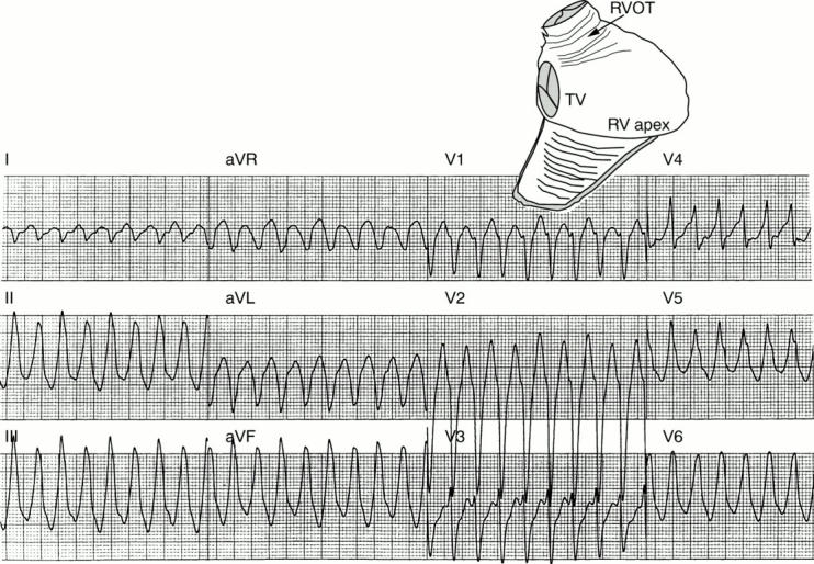
Figure 1: .

Idiopathic right ventricular outflow tract tachycardia. The 12 lead ECG shows tachycardia with a left bundle branch block, configuration and frontal plane axis directed inferiorly. The schematic at the upper right shows the right ventricle viewed from the right anterior oblique position with the free wall of the ventricle folded down. The location of the tachycardia in the right ventricular outflow tract (RVOT) is indicated with an arrow. TV, tricuspid valve; RV, right ventricle.
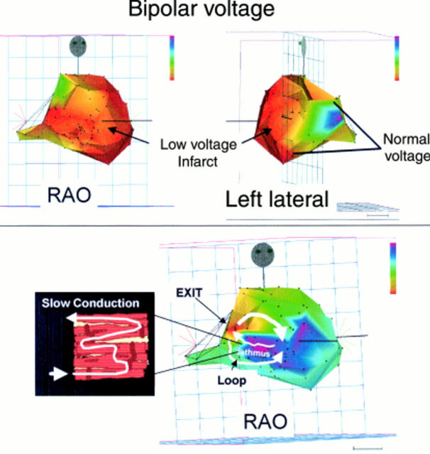
Figure 2: .
The mapping data are from a patient with VT late after anterior wall myocardial infarction. Mapping was performed using a system that plots the precise catheter position along with colour coded electrophysiologic information (CARTO Biosense Webster, Diamond Bar, California, USA). The top two panels show the left ventricle in right anterior oblique (RAO) and left lateral views. In this case, colours indicate the electrogram voltage, rather than timing. The lowest voltage regions are shown in red, progressing to greater voltage regions of yellow, green, blue, and purple. A large anteroapical infarction is indicated by the extensive low voltage, red region. The lower right panel shows the map of VT in the same patient. The ventricle is again shown in a right anterior oblique projection with the apex at the right and the base at the left hand side of the image. The colours indicate the activation sequence and arrows have been drawn to clarify the activation sequence of the circuit. The re-entry circuit is located in the septum. The wavefront starts at the red area (exit) near the base of the septum and splits into two loops that circle around the superior and inferior aspect of the septum toward the apex, re-entering an isthmus in the circuit that is proximal to the exit region. RF ablation in the isthmus abolished tachycardia. The mechanism of slow conduction through the infarct region that has been observed in previous histopathologic studies is illustrated schematically in the inset at lower left. Surviving myocyte bundles are separated by fibrous tissue that forces the wavefront to take a circuitous path through the region.
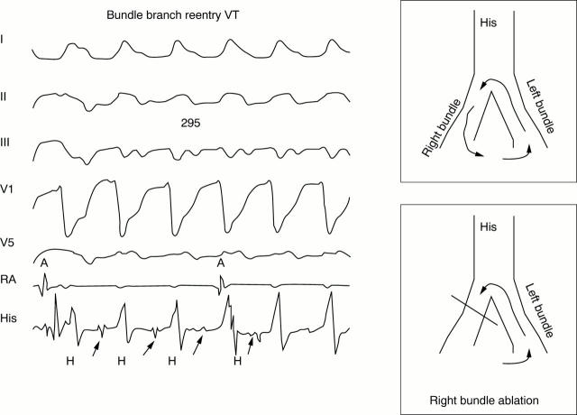
Figure 3: .
Bundle branch re-entry tachycardia. The left hand panel shows bundle branch re-entry tachycardia initiated in the electrophysiology laboratory. From the top are surface ECG leads and intracardiac recordings from the right atrium (RA) and His bundle position (His). VT has a left bundle branch block configuration and cycle length of 295 ms. Atrioventricular dissociation is evident in the right atrial recording (RA). A His bundle deflection (arrows) precedes each QRS indicating that the His-Purkinje system is closely linked to the tachycardia. The schematic in the right hand panels illustrates the mechanism. The wavefront circulates down the right bundle, through the interventricular septum, and up the left bundle (top panel). Ablation of the right bundle branch interrupts the circuit (bottom panel).
Selected References
These references are in PubMed. This may not be the complete list of references from this article.
- Blanck Z., Dhala A., Deshpande S., Sra J., Jazayeri M., Akhtar M. Bundle branch reentrant ventricular tachycardia: cumulative experience in 48 patients. J Cardiovasc Electrophysiol. 1993 Jun;4(3):253–262. doi: 10.1111/j.1540-8167.1993.tb01228.x. [DOI] [PubMed] [Google Scholar]
- Calkins H., Epstein A., Packer D., Arria A. M., Hummel J., Gilligan D. M., Trusso J., Carlson M., Luceri R., Kopelman H. Catheter ablation of ventricular tachycardia in patients with structural heart disease using cooled radiofrequency energy: results of a prospective multicenter study. Cooled RF Multi Center Investigators Group. J Am Coll Cardiol. 2000 Jun;35(7):1905–1914. doi: 10.1016/s0735-1097(00)00615-x. [DOI] [PubMed] [Google Scholar]
- Coggins D. L., Lee R. J., Sweeney J., Chein W. W., Van Hare G., Epstein L., Gonzalez R., Griffin J. C., Lesh M. D., Scheinman M. M. Radiofrequency catheter ablation as a cure for idiopathic tachycardia of both left and right ventricular origin. J Am Coll Cardiol. 1994 May;23(6):1333–1341. doi: 10.1016/0735-1097(94)90375-1. [DOI] [PubMed] [Google Scholar]
- Delacretaz E., Stevenson W. G., Ellison K. E., Maisel W. H., Friedman P. L. Mapping and radiofrequency catheter ablation of the three types of sustained monomorphic ventricular tachycardia in nonischemic heart disease. J Cardiovasc Electrophysiol. 2000 Jan;11(1):11–17. doi: 10.1111/j.1540-8167.2000.tb00728.x. [DOI] [PubMed] [Google Scholar]
- Ellison K. E., Friedman P. L., Ganz L. I., Stevenson W. G. Entrainment mapping and radiofrequency catheter ablation of ventricular tachycardia in right ventricular dysplasia. J Am Coll Cardiol. 1998 Sep;32(3):724–728. doi: 10.1016/s0735-1097(98)00292-7. [DOI] [PubMed] [Google Scholar]
- Ellison K. E., Stevenson W. G., Sweeney M. O., Lefroy D. C., Delacretaz E., Friedman P. L. Catheter ablation for hemodynamically unstable monomorphic ventricular tachycardia. J Cardiovasc Electrophysiol. 2000 Jan;11(1):41–44. doi: 10.1111/j.1540-8167.2000.tb00734.x. [DOI] [PubMed] [Google Scholar]
- Gonska B. D., Cao K., Schaumann A., Dorszewski A., von zur Mühlen F., Kreuzer H. Catheter ablation of ventricular tachycardia in 136 patients with coronary artery disease: results and long-term follow-up. J Am Coll Cardiol. 1994 Nov 15;24(6):1506–1514. doi: 10.1016/0735-1097(94)90147-3. [DOI] [PubMed] [Google Scholar]
- Lerman B. B., Stein K., Engelstein E. D., Battleman D. S., Lippman N., Bei D., Catanzaro D. Mechanism of repetitive monomorphic ventricular tachycardia. Circulation. 1995 Aug 1;92(3):421–429. doi: 10.1161/01.cir.92.3.421. [DOI] [PubMed] [Google Scholar]
- Marchlinski F. E., Callans D. J., Gottlieb C. D., Zado E. Linear ablation lesions for control of unmappable ventricular tachycardia in patients with ischemic and nonischemic cardiomyopathy. Circulation. 2000 Mar 21;101(11):1288–1296. doi: 10.1161/01.cir.101.11.1288. [DOI] [PubMed] [Google Scholar]
- Nakagawa H., Beckman K. J., McClelland J. H., Wang X., Arruda M., Santoro I., Hazlitt H. A., Abdalla I., Singh A., Gossinger H. Radiofrequency catheter ablation of idiopathic left ventricular tachycardia guided by a Purkinje potential. Circulation. 1993 Dec;88(6):2607–2617. doi: 10.1161/01.cir.88.6.2607. [DOI] [PubMed] [Google Scholar]
- Rodriguez L. M., Smeets J. L., Timmermans C., Wellens H. J. Predictors for successful ablation of right- and left-sided idiopathic ventricular tachycardia. Am J Cardiol. 1997 Feb 1;79(3):309–314. doi: 10.1016/s0002-9149(96)00753-9. [DOI] [PubMed] [Google Scholar]
- Rothman S. A., Hsia H. H., Cossú S. F., Chmielewski I. L., Buxton A. E., Miller J. M. Radiofrequency catheter ablation of postinfarction ventricular tachycardia: long-term success and the significance of inducible nonclinical arrhythmias. Circulation. 1997 Nov 18;96(10):3499–3508. doi: 10.1161/01.cir.96.10.3499. [DOI] [PubMed] [Google Scholar]
- Schalij M. J., van Rugge F. P., Siezenga M., van der Velde E. T. Endocardial activation mapping of ventricular tachycardia in patients : first application of a 32-site bipolar mapping electrode catheter. Circulation. 1998 Nov 17;98(20):2168–2179. doi: 10.1161/01.cir.98.20.2168. [DOI] [PubMed] [Google Scholar]
- Schilling R. J., Peters N. S., Davies D. W. Feasibility of a noncontact catheter for endocardial mapping of human ventricular tachycardia. Circulation. 1999 May 18;99(19):2543–2552. doi: 10.1161/01.cir.99.19.2543. [DOI] [PubMed] [Google Scholar]
- Sosa E., Scanavacca M., D'Avila A., Piccioni J., Sanchez O., Velarde J. L., Silva M., Reolão B. Endocardial and epicardial ablation guided by nonsurgical transthoracic epicardial mapping to treat recurrent ventricular tachycardia. J Cardiovasc Electrophysiol. 1998 Mar;9(3):229–239. doi: 10.1111/j.1540-8167.1998.tb00907.x. [DOI] [PubMed] [Google Scholar]
- Stevenson W. G., Delacretaz E., Friedman P. L., Ellison K. E. Identification and ablation of macroreentrant ventricular tachycardia with the CARTO electroanatomical mapping system. Pacing Clin Electrophysiol. 1998 Jul;21(7):1448–1456. doi: 10.1111/j.1540-8159.1998.tb00217.x. [DOI] [PubMed] [Google Scholar]
- Stevenson W. G., Friedman P. L., Kocovic D., Sager P. T., Saxon L. A., Pavri B. Radiofrequency catheter ablation of ventricular tachycardia after myocardial infarction. Circulation. 1998 Jul 28;98(4):308–314. doi: 10.1161/01.cir.98.4.308. [DOI] [PubMed] [Google Scholar]
- Strickberger S. A., Knight B. P., Michaud G. F., Pelosi F., Morady F. Mapping and ablation of ventricular tachycardia guided by virtual electrograms using a noncontact, computerized mapping system. J Am Coll Cardiol. 2000 Feb;35(2):414–421. doi: 10.1016/s0735-1097(99)00578-1. [DOI] [PubMed] [Google Scholar]
- Strickberger S. A., Man K. C., Daoud E. G., Goyal R., Brinkman K., Hasse C., Bogun F., Knight B. P., Weiss R., Bahu M. A prospective evaluation of catheter ablation of ventricular tachycardia as adjuvant therapy in patients with coronary artery disease and an implantable cardioverter-defibrillator. Circulation. 1997 Sep 2;96(5):1525–1531. doi: 10.1161/01.cir.96.5.1525. [DOI] [PubMed] [Google Scholar]
- de Bakker J. M., van Capelle F. J., Janse M. J., Tasseron S., Vermeulen J. T., de Jonge N., Lahpor J. R. Slow conduction in the infarcted human heart. 'Zigzag' course of activation. Circulation. 1993 Sep;88(3):915–926. doi: 10.1161/01.cir.88.3.915. [DOI] [PubMed] [Google Scholar]


