Full Text
The Full Text of this article is available as a PDF (104.5 KB).
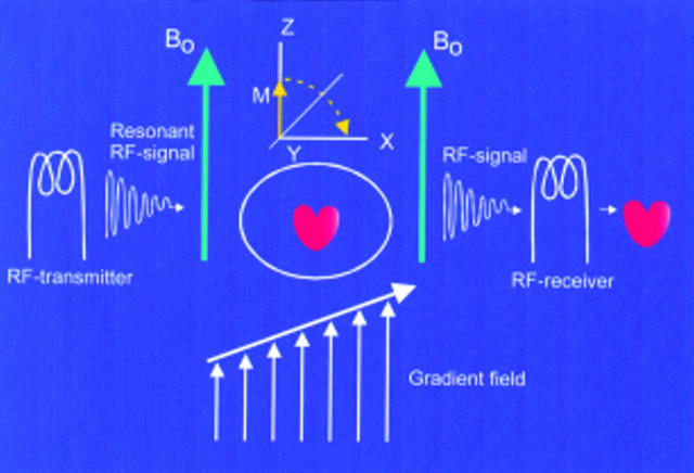
Figure 2 .
The patient (heart) is placed within a strong external magnetic field (Bo). The RF transmitter rotates the net tissue magnetisation in the transverse plane, and after termination, relaxation occurs which emits a signal detected by the RF receiver. The gradient coils produce a supplemental magnetic field gradient to allow precise location of the excited protons. The received signals have certain signal intensity (brightness) and location, both of which are processed to form the desired image.


