Abstract
OBJECTIVE—Contraction of longitudinal and subendocardial myocardial muscle fibres is reflected in descent of the atrioventricular (AV) plane. The aim was therefore to determine whether β blocker treatment with prolongation of diastole might result in improved function as reflected by AV plane movements in patients with chronic heart failure. DESIGN—Double blind, randomised, placebo controlled and open intervention study. SETTING—University hospital. PATIENTS—Patients with congestive heart failure: placebo controlled (n = 26) and an open protocol (n = 15). INTERVENTIONS—12 months of metoprolol treatment. MAIN OUTCOME MEASURES—Short axis and long axis echocardiography, invasive haemodynamics, radionuclide angiography. RESULTS—Recovery of systolic and diastolic function during metoprolol treatment was reflected by early changes in mean (SD) AV plane amplitude, from 5.3 (2.0)% to 7.1 (3.2)% and 7.8 (3.1)% (at 3 and 12 months, respectively; p < 0.05). In a multivariate analysis, only the change in AV plane amplitude by three months was independently associated with improvement in pulmonary capillary wedge pressure by six months (r = 0.80, p = 0.017). Change in AV plane amplitude by three months was also a better predictor of improvement in ejection fraction by 12 months (r = 0.78, p < 0.001) than changes in radionuclide ejection fraction by three months (r = 0.34, p = 0.049). CONCLUSIONS—Improvement in longitudinal contraction was closely associated with a decrease in left ventricular filling pressure during metoprolol treatment. This association was stronger than changes in short axis performance or radionuclide ejection fraction, emphasising the importance of AV plane motion for left ventricular filling and systolic performance in patients with heart failure. Keywords: diastolic function; metoprolol; dilated cardiomyopathy; echocardiography
Full Text
The Full Text of this article is available as a PDF (141.3 KB).
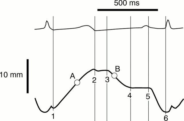
Figure 1 .
Schematic atrioventricular plane (AVP) curve and delineation of measured periods: (1) start of left ventricular contraction and ejection; (2) end of left ventricular contraction; (3) start of early rapid diastolic filling; (4) end of rapid filling, beginning of the diastasis; (5) end of diastasis, beginning of atrial contraction; (6) end of atrial contraction. (A) Maximum derivative of systolic ejection. (B) Maximum derivative of early rapid filling.
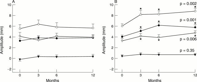
Figure 2 .
(A) Regional systolic amplitudes in patients on placebo. (B) Regional systolic amplitudes in patients on metoprolol treatment. Empty circles, left lateral atrioventricular plane (AVP); filled circles, septal AVP; empty triangles, posterior wall short axis; filled triangles, interventricular septum short axis. The p values of the analysis of variance test are shown to the right. *p < 0.05 v baseline investigation.
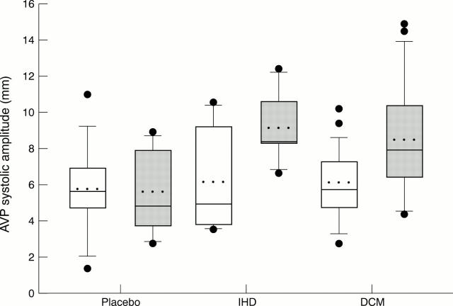
Figure 3 .
Box plots showing lateral atrioventricular plane (AVP) contraction amplitudes in patients on placebo (all dilated cardiomyopathy, n = 12), in patients with ischaemic cardiomyopathy (IHD, all on open metoprolol, n = 6), and in patients with dilated cardiomyopathy (DCM) treated with metoprolol (open treatment, n = 9; double blind treatment, n = 14). Baseline (white boxes), six months follow up (grey boxes). The boxes encompass the 25th to the 75th centile, and the whiskers the 10th to the 90th centile. Dots denote values outside the 10th and 90th centiles. Solid lines in the boxes denote median values and dotted lines denote mean values.
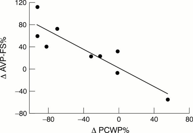
Figure 4 .
Linear correlation between systolic fractional shortening in the atrioventricular plane (AVP-FS) v pulmonary capillary wedge pressure (PCWP) in patients treated with metoprolol in the invasive substudy over six months. In a multivariate stepwise regression analysis, the AVP-FS was the only variable that was independently associated with the change in pulmonary capillary wedge pressure. ΔPCW % = −4.35 − 0.96(ΔAVP-FS %), r = 0.90, p = 0.001.
Figure 5 .
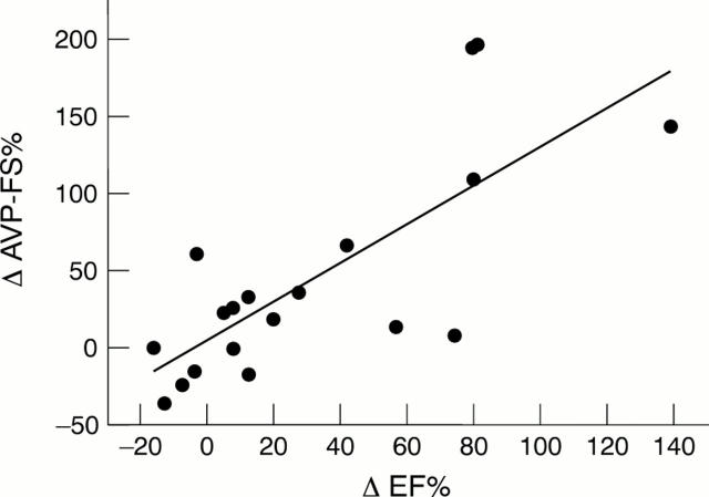
Linear correlation between systolic fractional shortening in the atrioventricular plane (AVP-FS) (over three months) versus change in radionuclide ejection fraction (EF) (over 12 months) in patients treated with metoprolol. In a multivariate stepwise regression analysis, the AVP-FS was the only echocardiographic variable that was independently associated with the future change in EF: ΔEF % = 12.3 + 0.46(ΔAVP-FS %), r = 0.76, p < 0.001.
Figure 6 .
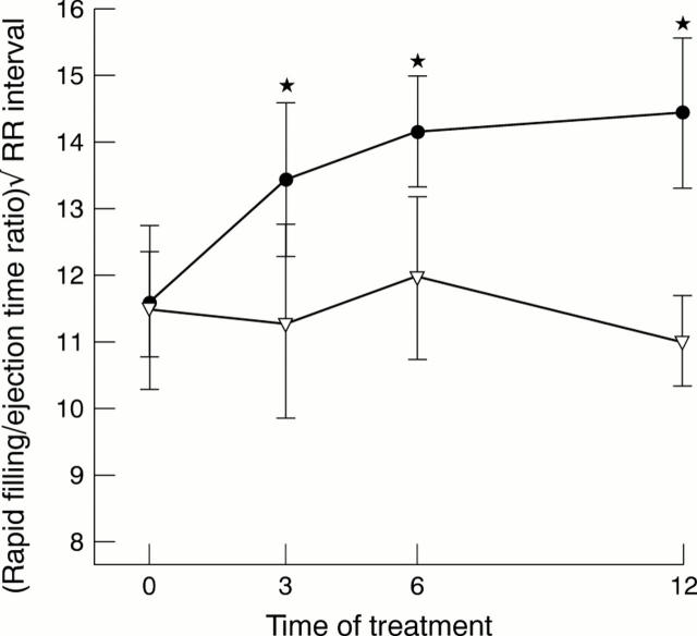
The atrioventricular plane (AVP) rapid filling time to ejection time ratio, corrected for heart rate by division with Q1Q2 interval. There was a significant prolongation of the rapid filling time to ejection time ratio in the metoprolol group, but no change in the placebo group. Filled circles, metoprolol; empty triangles, placebo. *p < 0.05 v baseline.
Selected References
These references are in PubMed. This may not be the complete list of references from this article.
- Alam M., Höglund C., Thorstrand C., Carlens P. Effects of exercise on the displacement of the atrioventricular plane in patients with coronary artery disease. A new echocardiographic method of detecting reversible myocardial ischaemia. Eur Heart J. 1991 Jul;12(7):760–765. [PubMed] [Google Scholar]
- Andersson B., Blomström-Lundqvist C., Hedner T., Waagstein F. Exercise hemodynamics and myocardial metabolism during long-term beta-adrenergic blockade in severe heart failure. J Am Coll Cardiol. 1991 Oct;18(4):1059–1066. doi: 10.1016/0735-1097(91)90767-4. [DOI] [PubMed] [Google Scholar]
- Andersson B., Caidahl K., di Lenarda A., Warren S. E., Goss F., Waldenström A., Persson S., Wallentin I., Hjalmarson A., Waagstein F. Changes in early and late diastolic filling patterns induced by long-term adrenergic beta-blockade in patients with idiopathic dilated cardiomyopathy. Circulation. 1996 Aug 15;94(4):673–682. doi: 10.1161/01.cir.94.4.673. [DOI] [PubMed] [Google Scholar]
- Andersson B., Hamm C., Persson S., Wikström G., Sinagra G., Hjalmarson A., Waagstein F. Improved exercise hemodynamic status in dilated cardiomyopathy after beta-adrenergic blockade treatment. J Am Coll Cardiol. 1994 May;23(6):1397–1404. doi: 10.1016/0735-1097(94)90383-2. [DOI] [PubMed] [Google Scholar]
- Andersson B., Lomsky M., Waagstein F. The link between acute haemodynamic adrenergic beta-blockade and long-term effects in patients with heart failure. A study on diastolic function, heart rate and myocardial metabolism following intravenous metoprolol. Eur Heart J. 1993 Oct;14(10):1375–1385. doi: 10.1093/eurheartj/14.10.1375. [DOI] [PubMed] [Google Scholar]
- Andersson B., Strömblad S. O., Lomsky M., Waagstein F. Heart rate dependency of cardiac performance in heart failure patients treated with metoprolol. Eur Heart J. 1999 Apr;20(8):575–583. doi: 10.1053/euhj.1998.1315. [DOI] [PubMed] [Google Scholar]
- Beau S. L., Saffitz J. E. Transmural heterogeneity of norepinephrine uptake in failing human hearts. J Am Coll Cardiol. 1994 Mar 1;23(3):579–585. doi: 10.1016/0735-1097(94)90739-0. [DOI] [PubMed] [Google Scholar]
- Beau S. L., Tolley T. K., Saffitz J. E. Heterogeneous transmural distribution of beta-adrenergic receptor subtypes in failing human hearts. Circulation. 1993 Dec;88(6):2501–2509. doi: 10.1161/01.cir.88.6.2501. [DOI] [PubMed] [Google Scholar]
- Birkeland S., Westby J., Grong K., Lekven J. Effect of afterload and beta-adrenergic blockade on nonischemic myocardial contraction pattern. Am J Physiol. 1992 Dec;263(6 Pt 2):H1716–H1723. doi: 10.1152/ajpheart.1992.263.6.H1716. [DOI] [PubMed] [Google Scholar]
- Böhm M., Deutsch H. J., Hartmann D., Rosée K. L., Stäblein A. Improvement of postreceptor events by metoprolol treatment in patients with chronic heart failure. J Am Coll Cardiol. 1997 Oct;30(4):992–996. doi: 10.1016/s0735-1097(97)00248-9. [DOI] [PubMed] [Google Scholar]
- Garcia M. J., Rodriguez L., Ares M., Griffin B. P., Thomas J. D., Klein A. L. Differentiation of constrictive pericarditis from restrictive cardiomyopathy: assessment of left ventricular diastolic velocities in longitudinal axis by Doppler tissue imaging. J Am Coll Cardiol. 1996 Jan;27(1):108–114. doi: 10.1016/0735-1097(95)00434-3. [DOI] [PubMed] [Google Scholar]
- Gwathmey J. K., Kim C. S., Hajjar R. J., Khan F., DiSalvo T. G., Matsumori A., Bristow M. R. Cellular and molecular remodeling in a heart failure model treated with the beta-blocker carteolol. Am J Physiol. 1999 May;276(5 Pt 2):H1678–H1690. doi: 10.1152/ajpheart.1999.276.5.H1678. [DOI] [PubMed] [Google Scholar]
- Heilbrunn S. M., Shah P., Bristow M. R., Valantine H. A., Ginsburg R., Fowler M. B. Increased beta-receptor density and improved hemodynamic response to catecholamine stimulation during long-term metoprolol therapy in heart failure from dilated cardiomyopathy. Circulation. 1989 Mar;79(3):483–490. doi: 10.1161/01.cir.79.3.483. [DOI] [PubMed] [Google Scholar]
- Henein M. Y., Priestley K., Davarashvili T., Buller N., Gibson D. G. Early changes in left ventricular subendocardial function after successful coronary angioplasty. Br Heart J. 1993 Jun;69(6):501–506. doi: 10.1136/hrt.69.6.501. [DOI] [PMC free article] [PubMed] [Google Scholar]
- Quaife R. A., Gilbert E. M., Christian P. E., Datz F. L., Mealey P. C., Volkman K., Olsen S. L., Bristow M. R. Effects of carvedilol on systolic and diastolic left ventricular performance in idiopathic dilated cardiomyopathy or ischemic cardiomyopathy. Am J Cardiol. 1996 Oct 1;78(7):779–784. doi: 10.1016/s0002-9149(96)00420-1. [DOI] [PubMed] [Google Scholar]
- Rogers W. J., Jr, Shapiro E. P., Weiss J. L., Buchalter M. B., Rademakers F. E., Weisfeldt M. L., Zerhouni E. A. Quantification of and correction for left ventricular systolic long-axis shortening by magnetic resonance tissue tagging and slice isolation. Circulation. 1991 Aug;84(2):721–731. doi: 10.1161/01.cir.84.2.721. [DOI] [PubMed] [Google Scholar]
- Sahn D. J., DeMaria A., Kisslo J., Weyman A. Recommendations regarding quantitation in M-mode echocardiography: results of a survey of echocardiographic measurements. Circulation. 1978 Dec;58(6):1072–1083. doi: 10.1161/01.cir.58.6.1072. [DOI] [PubMed] [Google Scholar]
- Tsakiris A. G., Gordon D. A., Padiyar R., Fréchette D., Labrosse C. The role of displacement of the mitral annulus in left atrial filling and emptying in the intact dog. Can J Physiol Pharmacol. 1978 Jun;56(3):447–457. doi: 10.1139/y78-067. [DOI] [PubMed] [Google Scholar]
- Waagstein F., Bristow M. R., Swedberg K., Camerini F., Fowler M. B., Silver M. A., Gilbert E. M., Johnson M. R., Goss F. G., Hjalmarson A. Beneficial effects of metoprolol in idiopathic dilated cardiomyopathy. Metoprolol in Dilated Cardiomyopathy (MDC) Trial Study Group. Lancet. 1993 Dec 11;342(8885):1441–1446. doi: 10.1016/0140-6736(93)92930-r. [DOI] [PubMed] [Google Scholar]
- Waagstein F., Caidahl K., Wallentin I., Bergh C. H., Hjalmarson A. Long-term beta-blockade in dilated cardiomyopathy. Effects of short- and long-term metoprolol treatment followed by withdrawal and readministration of metoprolol. Circulation. 1989 Sep;80(3):551–563. doi: 10.1161/01.cir.80.3.551. [DOI] [PubMed] [Google Scholar]
- Willenheimer R., Cline C., Erhardt L., Israelsson B. Left ventricular atrioventricular plane displacement: an echocardiographic technique for rapid assessment of prognosis in heart failure. Heart. 1997 Sep;78(3):230–236. doi: 10.1136/hrt.78.3.230. [DOI] [PMC free article] [PubMed] [Google Scholar]


