

Abstract
OBJECTIVE—To determine the pulmonary venous flow velocity (PVFV) values in a large normal population. DESIGN—Prospective study in consecutive individuals. SETTING—University hospital. METHODS—Among 404 normal individuals, the flow velocity pattern in the right upper pulmonary vein was recorded in 315 subjects using transthoracic echocardiography, and in both upper pulmonary veins in 100 subjects using transoesophageal echocardiography. Subjects were divided into five age groups. The PVFV values were compared between transthoracic and transoesophageal echocardiography within the age groups, and intraindividually between the right and left upper pulmonary veins in transoesophageal echocardiography. RESULTS—Normal PVFV values for the right upper pulmonary vein in transthoracic and transoesophageal echocardiography are presented. The duration of flow reversal at atrial contraction was overestimated using transthoracic echocardiography (mean (SD): 96 (21) ms in transoesophageal echocardiography, 120 (28) ms in transthoracic echocardiography, p < 0.0001). Systolic to diastolic peak flow velocity ratio (S:D) increased earlier with advancing age with transoesophageal echocardiography than with transthoracic echocardiography. Similar results were found for the corresponding time-velocity integrals. Data from the left and right upper pulmonary veins differed with respect to onset and deceleration of flow velocities, but not for flow durations or peak velocities. CONCLUSIONS—Normal PVFV values generally show a wide range. The data presented will be of value in assessing left ventricular diastolic function and mitral regurgitation using the PVFV pattern. Keywords: pulmonary venous flow velocity; Doppler echocardiography; mitral regurgitation
Full Text
The Full Text of this article is available as a PDF (199.9 KB).
Figure 1 .

(A) Transoesophageal Doppler echocardiographic recording of right upper pulmonary venous flow in a 67 year old man. (B) Illustration of the flow velocity variables measured in each pulmonary vein. Explanations of abbreviations are given in the box.
Figure 2 .
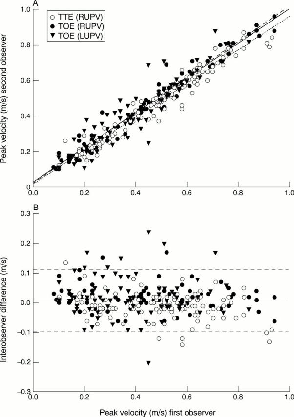
(A) Correlation between first and second observers' velocity measurements (systolic, diastolic, and reversed flow at atrial contraction) in transthoracic and transoesophageal echocardiography. (B) Bland-Altmann plot showing the interobserver variability of pulmonary venous flow velocity measurements in transthoracic and transoesophageal echocardiography. The solid line indicates the mean and the dashed lines the 95% confidence intervals of the interobserver difference of all flow velocity measurements. LUPV, left upper pulmonary vein; RUPV, right upper pulmonary vein; TOE, transoesophageal echocardiography; TTE, transthoracic echocardiography.
Figure 3 .

Relation between age and the systolic to diastolic peak flow velocity ratio (S:D). There was a significant increase in S:D with advancing age in both transthoracic and transoesophageal echocardiography. However, this increase was less pronounced in transthoracic echocardiography. The dotted arrows indicate the age at which the regression lines exceed S:D = 1 (that is, 41 years in transthoracic and 32 years in transoesophageal echocardiography). Note that there is an abundant scatter of normal S:D values in all age groups. LUPV, left upper pulmonary vein; RUPV, right upper pulmonary vein; TOE, transoesophageal echocardiography; TTE, transthoracic echocardiography.
Selected References
These references are in PubMed. This may not be the complete list of references from this article.
- Brun P., Tribouilloy C., Duval A. M., Iserin L., Meguira A., Pelle G., Dubois-Rande J. L. Left ventricular flow propagation during early filling is related to wall relaxation: a color M-mode Doppler analysis. J Am Coll Cardiol. 1992 Aug;20(2):420–432. doi: 10.1016/0735-1097(92)90112-z. [DOI] [PubMed] [Google Scholar]
- Castello R., Pearson A. C., Lenzen P., Labovitz A. J. Evaluation of pulmonary venous flow by transesophageal echocardiography in subjects with a normal heart: comparison with transthoracic echocardiography. J Am Coll Cardiol. 1991 Jul;18(1):65–71. doi: 10.1016/s0735-1097(10)80219-0. [DOI] [PubMed] [Google Scholar]
- Devereux R. B., Lutas E. M., Casale P. N., Kligfield P., Eisenberg R. R., Hammond I. W., Miller D. H., Reis G., Alderman M. H., Laragh J. H. Standardization of M-mode echocardiographic left ventricular anatomic measurements. J Am Coll Cardiol. 1984 Dec;4(6):1222–1230. doi: 10.1016/s0735-1097(84)80141-2. [DOI] [PubMed] [Google Scholar]
- Gentile F., Mantero A., Lippolis A., Ornaghi M., Azzollini M., Barbier P., Beretta L., Casazza F., Corno R., Faletra F. Pulmonary venous flow velocity patterns in 143 normal subjects aged 20 to 80 years old. An echo 2D colour Doppler cooperative study. Eur Heart J. 1997 Jan;18(1):148–164. doi: 10.1093/oxfordjournals.eurheartj.a015097. [DOI] [PubMed] [Google Scholar]
- Hoit B. D., Shao Y., Gabel M., Walsh R. A. Influence of loading conditions and contractile state on pulmonary venous flow. Validation of Doppler velocimetry. Circulation. 1992 Aug;86(2):651–659. doi: 10.1161/01.cir.86.2.651. [DOI] [PubMed] [Google Scholar]
- Jensen J. L., Williams F. E., Beilby B. J., Johnson B. L., Miller L. K., Ginter T. L., Tomaselli-Martin G., Appleton C. P. Feasibility of obtaining pulmonary venous flow velocity in cardiac patients using transthoracic pulsed wave Doppler technique. J Am Soc Echocardiogr. 1997 Jan-Feb;10(1):60–66. doi: 10.1016/s0894-7317(97)80033-8. [DOI] [PubMed] [Google Scholar]
- Klein A. L., Obarski T. P., Stewart W. J., Casale P. N., Pearce G. L., Husbands K., Cosgrove D. M., Salcedo E. E. Transesophageal Doppler echocardiography of pulmonary venous flow: a new marker of mitral regurgitation severity. J Am Coll Cardiol. 1991 Aug;18(2):518–526. doi: 10.1016/0735-1097(91)90609-d. [DOI] [PubMed] [Google Scholar]
- Klein A. L., Tajik A. J. Doppler assessment of pulmonary venous flow in healthy subjects and in patients with heart disease. J Am Soc Echocardiogr. 1991 Jul-Aug;4(4):379–392. doi: 10.1016/s0894-7317(14)80448-3. [DOI] [PubMed] [Google Scholar]
- Mayet J., More R. S., Sutton G. C. Anticoagulation for cardioversion of atrial arrhythmias. Eur Heart J. 1998 Apr;19(4):548–552. doi: 10.1053/euhj.1997.0509. [DOI] [PubMed] [Google Scholar]
- Nagueh S. F., Middleton K. J., Kopelen H. A., Zoghbi W. A., Quiñones M. A. Doppler tissue imaging: a noninvasive technique for evaluation of left ventricular relaxation and estimation of filling pressures. J Am Coll Cardiol. 1997 Nov 15;30(6):1527–1533. doi: 10.1016/s0735-1097(97)00344-6. [DOI] [PubMed] [Google Scholar]
- Nakatani S., Yoshitomi H., Wada K., Beppu S., Nagata S., Miyatake K. Noninvasive estimation of left ventricular end-diastolic pressure using transthoracic Doppler-determined pulmonary venous atrial flow reversal. Am J Cardiol. 1994 May 15;73(13):1017–1018. doi: 10.1016/0002-9149(94)90162-7. [DOI] [PubMed] [Google Scholar]
- Nishimura R. A., Abel M. D., Hatle L. K., Tajik A. J. Relation of pulmonary vein to mitral flow velocities by transesophageal Doppler echocardiography. Effect of different loading conditions. Circulation. 1990 May;81(5):1488–1497. doi: 10.1161/01.cir.81.5.1488. [DOI] [PubMed] [Google Scholar]
- Oki T., Tabata T., Yamada H., Wakatsuki T., Shinohara H., Nishikado A., Iuchi A., Fukuda N., Ito S. Clinical application of pulsed Doppler tissue imaging for assessing abnormal left ventricular relaxation. Am J Cardiol. 1997 Apr 1;79(7):921–928. doi: 10.1016/s0002-9149(97)00015-5. [DOI] [PubMed] [Google Scholar]
- Rossvoll O., Hatle L. K. Pulmonary venous flow velocities recorded by transthoracic Doppler ultrasound: relation to left ventricular diastolic pressures. J Am Coll Cardiol. 1993 Jun;21(7):1687–1696. doi: 10.1016/0735-1097(93)90388-h. [DOI] [PubMed] [Google Scholar]
- Seiler C., Aeschbacher B. C., Meier B. Quantitation of mitral regurgitation using the systolic/diastolic pulmonary venous flow velocity ratio. J Am Coll Cardiol. 1998 May;31(6):1383–1390. doi: 10.1016/s0735-1097(98)00090-4. [DOI] [PubMed] [Google Scholar]
- Steen T., Voss B. M., Smiseth O. A. Influence of heart rate and left atrial pressure on pulmonary venous flow pattern in dogs. Am J Physiol. 1994 Jun;266(6 Pt 2):H2296–H2302. doi: 10.1152/ajpheart.1994.266.6.H2296. [DOI] [PubMed] [Google Scholar]