Abstract
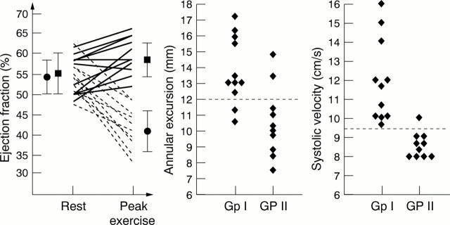
OBJECTIVE—To identify variables that could be applied at rest to diagnose subclinical ventricular dysfunction in asymptomatic patients with severe aortic regurgitation. DESIGN—Cross sectional study. PATIENTS—Left ventricular long axis contraction was studied using tissue Doppler and M mode echocardiography in 21 patients with no symptoms (New York Heart Association (NYHA) functional class ⩽ 2a) but severe aortic regurgitation (jet area/left ventricular outflow tract area > 40%). MAIN OUTCOME MEASURES—Left ventricular ejection fraction (LVEF) at baseline and peak exercise (Weber protocol), cardiopulmonary function, and left ventricular long axis function at rest (peak systolic velocity and excursion of the mitral annulus). RESULTS—In 11 patients, ejection fraction increased or did not change (from mean (SD) 55 (5)% to 58 (4)%, p < 0.05) (group I); in 10 patients it decreased by > 5% (from 54 (4)% to 42 (5)%, p < 0.001) (group II). Exercise ejection fraction was < 50% in all patients in group II. At rest, there were no differences between the groups in ejection fraction, left ventricular diameter indices, wall stress, and short axis contraction. However, patients in group II had reduced long axis contraction compared with group I: peak systolic velocity 8.6 (0.6) v 11.9 (2.2) cm/s (p < 0.001); excursion 11 (2) v 14 (2) mm (p < 0.01). A resting velocity of < 9.5 cm/s was the best indicator of poor exercise tolerance (sensitivity 90%, specificity 100%). CONCLUSIONS—Markers of reduced long axis contraction may provide simple and reliable indices of subclinical left ventricular dysfunction in asymptomatic patients with severe aortic regurgitation. Keywords: aortic regurgitation; long axis function; tissue Doppler echocardiography; exercise echocardiography
Full Text
The Full Text of this article is available as a PDF (161.4 KB).
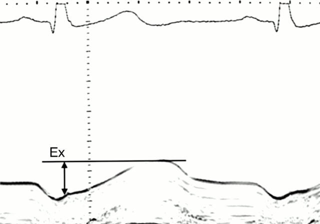
Figure 1 .
M mode trace of medial mitral annular motion recorded from an apical window. Ex, systolic excursion. Scale, 1 cm per division.
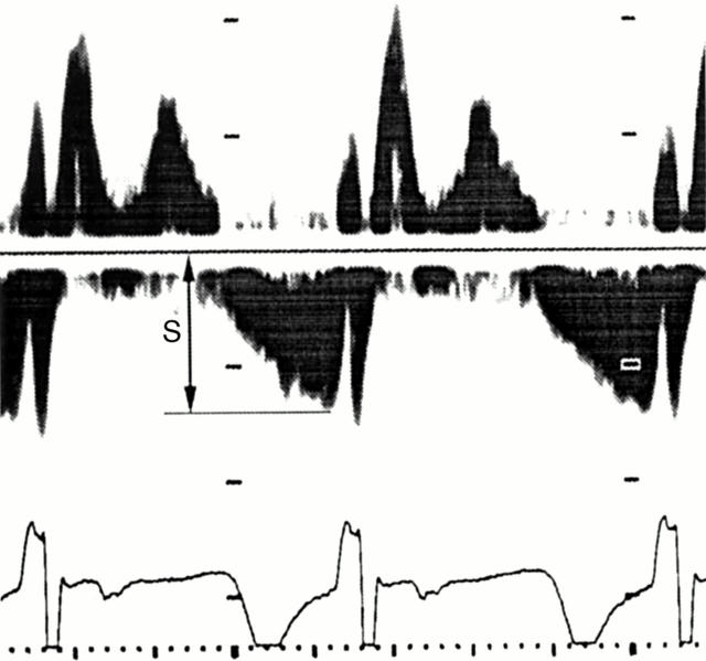
Figure 2 .
Pulsed wave tissue Doppler echocardiography of medial mitral annular motion. S, peak systolic velocity. Scale, 10 cm/s per division.
Figure 3 .
Left: individual rest and peak exercise ejection fraction values for patients with good exercise responses (group I) (solid lines) and poor exercise responses (group II) (dashed lines); in group I mean ejection fraction increased from 55% to 58% (solid squares), and in group II it decreased from 54% to 42% (solid circles). Middle: Distributions of individual values of systolic excursion and peak systolic velocity (right) of medial mitral annular motion, in patients with good exercise response (group I) and poor exercise response (group II). The dotted lines represent the best discriminant cut off values obtained in the analyses of sensitivity and specificity.
Selected References
These references are in PubMed. This may not be the complete list of references from this article.
- Assmann P. E., Slager C. J., van der Borden S. G., Dreysse S. T., Tijssen J. G., Sutherland G. R., Roelandt J. R. Quantitative echocardiographic analysis of global and regional left ventricular function: a problem revisited. J Am Soc Echocardiogr. 1990 Nov-Dec;3(6):478–487. doi: 10.1016/s0894-7317(14)80364-7. [DOI] [PubMed] [Google Scholar]
- Bland J. M., Altman D. G. Statistical methods for assessing agreement between two methods of clinical measurement. Lancet. 1986 Feb 8;1(8476):307–310. [PubMed] [Google Scholar]
- Bonow R. O., Lakatos E., Maron B. J., Epstein S. E. Serial long-term assessment of the natural history of asymptomatic patients with chronic aortic regurgitation and normal left ventricular systolic function. Circulation. 1991 Oct;84(4):1625–1635. doi: 10.1161/01.cir.84.4.1625. [DOI] [PubMed] [Google Scholar]
- Bonow R. O., Rosing D. R., Maron B. J., McIntosh C. L., Jones M., Bacharach S. L., Green M. V., Clark R. E., Epstein S. E. Reversal of left ventricular dysfunction after aortic valve replacement for chronic aortic regurgitation: influence of duration of preoperative left ventricular dysfunction. Circulation. 1984 Oct;70(4):570–579. doi: 10.1161/01.cir.70.4.570. [DOI] [PubMed] [Google Scholar]
- Corrao S., Paterna S., Arnone S., Costa R., Amato V., Amico G., Scaglione R., Licata G. Two-dimensional echocardiographic evaluation of left ventricular ejection fraction by the ellipsoid single-plane algorithm: a reliable method for assessing low or very low ejection fraction values? Cardiology. 1995;86(6):503–507. doi: 10.1159/000176930. [DOI] [PubMed] [Google Scholar]
- Fast J., Jacobs S. Limits of reproducibility of cross-sectional echocardiographic measurement of left ventricular ejection fraction. Int J Cardiol. 1990 Jul;28(1):67–72. doi: 10.1016/0167-5273(90)90010-3. [DOI] [PubMed] [Google Scholar]
- Garcia M. J., Rodriguez L., Ares M., Griffin B. P., Klein A. L., Stewart W. J., Thomas J. D. Myocardial wall velocity assessment by pulsed Doppler tissue imaging: characteristic findings in normal subjects. Am Heart J. 1996 Sep;132(3):648–656. doi: 10.1016/s0002-8703(96)90251-3. [DOI] [PubMed] [Google Scholar]
- Gorcsan J., 3rd, Gulati V. K., Mandarino W. A., Katz W. E. Color-coded measures of myocardial velocity throughout the cardiac cycle by tissue Doppler imaging to quantify regional left ventricular function. Am Heart J. 1996 Jun;131(6):1203–1213. doi: 10.1016/s0002-8703(96)90097-6. [DOI] [PubMed] [Google Scholar]
- Gordon E. P., Schnittger I., Fitzgerald P. J., Williams P., Popp R. L. Reproducibility of left ventricular volumes by two-dimensional echocardiography. J Am Coll Cardiol. 1983 Sep;2(3):506–513. doi: 10.1016/s0735-1097(83)80278-2. [DOI] [PubMed] [Google Scholar]
- Greenbaum R. A., Ho S. Y., Gibson D. G., Becker A. E., Anderson R. H. Left ventricular fibre architecture in man. Br Heart J. 1981 Mar;45(3):248–263. doi: 10.1136/hrt.45.3.248. [DOI] [PMC free article] [PubMed] [Google Scholar]
- Grossman W., Jones D., McLaurin L. P. Wall stress and patterns of hypertrophy in the human left ventricle. J Clin Invest. 1975 Jul;56(1):56–64. doi: 10.1172/JCI108079. [DOI] [PMC free article] [PubMed] [Google Scholar]
- Gulati V. K., Katz W. E., Follansbee W. P., Gorcsan J., 3rd Mitral annular descent velocity by tissue Doppler echocardiography as an index of global left ventricular function. Am J Cardiol. 1996 May 1;77(11):979–984. doi: 10.1016/s0002-9149(96)00033-1. [DOI] [PubMed] [Google Scholar]
- Henry W. L., Bonow R. O., Rosing D. R., Epstein S. E. Observations on the optimum time for operative intervention for aortic regurgitation. II. Serial echocardiographic evaluation of asymptomatic patients. Circulation. 1980 Mar;61(3):484–492. doi: 10.1161/01.cir.61.3.484. [DOI] [PubMed] [Google Scholar]
- Ibrahim M., O'Kane H., Cleland J., Gladstone D., Sarsam M., Patterson C. The St. Jude Medical prosthesis. A thirteen-year experience. J Thorac Cardiovasc Surg. 1994 Aug;108(2):221–230. [PubMed] [Google Scholar]
- Isaaz K., Munoz del Romeral L., Lee E., Schiller N. B. Quantitation of the motion of the cardiac base in normal subjects by Doppler echocardiography. J Am Soc Echocardiogr. 1993 Mar-Apr;6(2):166–176. doi: 10.1016/s0894-7317(14)80487-2. [DOI] [PubMed] [Google Scholar]
- Jones C. J., Raposo L., Gibson D. G. Functional importance of the long axis dynamics of the human left ventricle. Br Heart J. 1990 Apr;63(4):215–220. doi: 10.1136/hrt.63.4.215. [DOI] [PMC free article] [PubMed] [Google Scholar]
- Klodas E., Enriquez-Sarano M., Tajik A. J., Mullany C. J., Bailey K. R., Seward J. B. Optimizing timing of surgical correction in patients with severe aortic regurgitation: role of symptoms. J Am Coll Cardiol. 1997 Sep;30(3):746–752. doi: 10.1016/s0735-1097(97)00205-2. [DOI] [PubMed] [Google Scholar]
- Klodas E., Enriquez-Sarano M., Tajik A. J., Mullany C. J., Bailey K. R., Seward J. B. Surgery for aortic regurgitation in women. Contrasting indications and outcomes compared with men. Circulation. 1996 Nov 15;94(10):2472–2478. doi: 10.1161/01.cir.94.10.2472. [DOI] [PubMed] [Google Scholar]
- Lewis N. P., Banning A. P., Cooper J. P., Sundar A. S., Facey P. E., Evans W. D., Henderson A. H. Impaired matching of perfusion and ventilation in heart failure detected by 133xenon. Basic Res Cardiol. 1996;91 (Suppl 1):45–49. doi: 10.1007/BF00810523. [DOI] [PubMed] [Google Scholar]
- Magid N. M., Wallerson D. C., Borer J. S., Mukherjee A., Young M. S., Devereux R. B., Carter J. N. Left ventricular diastolic and systolic performance during chronic experimental aortic regurgitation. Am J Physiol. 1992 Jul;263(1 Pt 2):H226–H233. doi: 10.1152/ajpheart.1992.263.1.H226. [DOI] [PubMed] [Google Scholar]
- Massie B. M., Kramer B. L., Loge D., Topic N., Greenberg B. H., Cheitlin M. D., Bristow J. D., Byrd R. C. Ejection fraction response to supine exercise in asymptomatic aortic regurgitation: relation to simultaneous hemodynamic measurements. J Am Coll Cardiol. 1985 Apr;5(4):847–855. doi: 10.1016/s0735-1097(85)80422-8. [DOI] [PubMed] [Google Scholar]
- Nakagawa M., Shirato K., Ohyama T., Sakuma M., Takishima T. Left ventricular end-systolic stress-volume index ratio in aortic and mitral regurgitation with normal ejection fraction. Am Heart J. 1990 Oct;120(4):892–901. doi: 10.1016/0002-8703(90)90207-e. [DOI] [PubMed] [Google Scholar]
- Oh J. K., Hatle L. K., Sinak L. J., Seward J. B., Tajik A. J. Characteristic Doppler echocardiographic pattern of mitral inflow velocity in severe aortic regurgitation. J Am Coll Cardiol. 1989 Dec;14(7):1712–1717. doi: 10.1016/0735-1097(89)90021-1. [DOI] [PubMed] [Google Scholar]
- Opasich C., Pinna G. D., Bobbio M., Sisti M., Demichelis B., Febo O., Forni G., Riccardi R., Riccardi P. G., Capomolla S. Peak exercise oxygen consumption in chronic heart failure: toward efficient use in the individual patient. J Am Coll Cardiol. 1998 Mar 15;31(4):766–775. doi: 10.1016/s0735-1097(98)00002-3. [DOI] [PubMed] [Google Scholar]
- Pai R. G., Bodenheimer M. M., Pai S. M., Koss J. H., Adamick R. D. Usefulness of systolic excursion of the mitral anulus as an index of left ventricular systolic function. Am J Cardiol. 1991 Jan 15;67(2):222–224. doi: 10.1016/0002-9149(91)90453-r. [DOI] [PubMed] [Google Scholar]
- Palka P., Lange A., Fleming A. D., Fenn L. N., Bouki K. P., Shaw T. R., Fox K. A., McDicken W. N., Sutherland G. R. Age-related transmural peak mean velocities and peak velocity gradients by Doppler myocardial imaging in normal subjects. Eur Heart J. 1996 Jun;17(6):940–950. doi: 10.1093/oxfordjournals.eurheartj.a014977. [DOI] [PubMed] [Google Scholar]
- Park S. H., Shub C., Nobrega T. P., Bailey K. R., Seward J. B. Two-dimensional echocardiographic calculation of left ventricular mass as recommended by the American Society of Echocardiography: correlation with autopsy and M-mode echocardiography. J Am Soc Echocardiogr. 1996 Mar-Apr;9(2):119–128. doi: 10.1016/s0894-7317(96)90019-x. [DOI] [PubMed] [Google Scholar]
- Percy R. F., Miller A. B., Conetta D. A. Usefulness of left ventricular wall stress at rest and after exercise for outcome prediction in asymptomatic aortic regurgitation. Am Heart J. 1993 Jan;125(1):151–155. doi: 10.1016/0002-8703(93)90068-k. [DOI] [PubMed] [Google Scholar]
- Schiller N. B., Shah P. M., Crawford M., DeMaria A., Devereux R., Feigenbaum H., Gutgesell H., Reichek N., Sahn D., Schnittger I. Recommendations for quantitation of the left ventricle by two-dimensional echocardiography. American Society of Echocardiography Committee on Standards, Subcommittee on Quantitation of Two-Dimensional Echocardiograms. J Am Soc Echocardiogr. 1989 Sep-Oct;2(5):358–367. doi: 10.1016/s0894-7317(89)80014-8. [DOI] [PubMed] [Google Scholar]
- Scriven A. J., Lipkin D. P., Fox K. M., Poole-Wilson P. A. Maximal oxygen uptake in severe aortic regurgitation: a different view of left ventricular function. Am Heart J. 1990 Oct;120(4):902–909. doi: 10.1016/0002-8703(90)90208-f. [DOI] [PubMed] [Google Scholar]
- Shen W. F., Roubin G. S., Choong C. Y., Hutton B. F., Harris P. J., Fletcher P. J., Kelly D. T. Evaluation of relationship between myocardial contractile state and left ventricular function in patients with aortic regurgitation. Circulation. 1985 Jan;71(1):31–38. doi: 10.1161/01.cir.71.1.31. [DOI] [PubMed] [Google Scholar]
- Siemienczuk D., Greenberg B., Morris C., Massie B., Wilson R. A., Topic N., Bristow J. D., Cheitlin M. Chronic aortic insufficiency: factors associated with progression to aortic valve replacement. Ann Intern Med. 1989 Apr 15;110(8):587–592. doi: 10.7326/0003-4819-110-8-587. [DOI] [PubMed] [Google Scholar]
- Simonson J. S., Schiller N. B. Descent of the base of the left ventricle: an echocardiographic index of left ventricular function. J Am Soc Echocardiogr. 1989 Jan-Feb;2(1):25–35. doi: 10.1016/s0894-7317(89)80026-4. [DOI] [PubMed] [Google Scholar]
- Sohn D. W., Chai I. H., Lee D. J., Kim H. C., Kim H. S., Oh B. H., Lee M. M., Park Y. B., Choi Y. S., Seo J. D. Assessment of mitral annulus velocity by Doppler tissue imaging in the evaluation of left ventricular diastolic function. J Am Coll Cardiol. 1997 Aug;30(2):474–480. doi: 10.1016/s0735-1097(97)88335-0. [DOI] [PubMed] [Google Scholar]
- Villari B., Campbell S. E., Hess O. M., Mall G., Vassalli G., Weber K. T., Krayenbuehl H. P. Influence of collagen network on left ventricular systolic and diastolic function in aortic valve disease. J Am Coll Cardiol. 1993 Nov 1;22(5):1477–1484. doi: 10.1016/0735-1097(93)90560-n. [DOI] [PubMed] [Google Scholar]
- Vinereanu D., Khokhar A., Fraser A. G. Reproducibility of pulsed wave tissue Doppler echocardiography. J Am Soc Echocardiogr. 1999 Jun;12(6):492–499. doi: 10.1016/s0894-7317(99)70086-6. [DOI] [PubMed] [Google Scholar]


