Abstract
OBJECTIVE—To apply three dimensional echocardiography to describe the geometric profile of the Amplatzer and Cardioseal occluders after deployment for closure of atrial septal defect. METHODS—20 patients (mean (SD) age, 14 (5) years) were enrolled for transcatheter closure of a secundum atrial septal defect with the Amplatzer occluder (10) or with the Cardioseal occluder (10). The two populations were matched for the stretched diameter of the defect (mean 18 (6) mm). The profile of the two occluders was examined. RESULTS—Transoesophageal echocardiography did not show any residual shunts after Amplatzer occluder deployment, whereas three patients had a small residual leak after Cardioseal deployment. One patient had transient atrioventricular block with the Amplatzer device. The mean surface area of the Amplatzer occluder was 6.9 (2) cm2, and that of the Cardioseal device 5.4 (3) cm2 (p = 0.03). The mean volume of the Amplatzer occluder was 9.2 (1) cm3, while that of the Cardioseal occluder was 3.5 (1) cm3 (p < 0.0001). From the three dimensional views, the Cardioseal occluder looked like a flat square after deployment whereas the Amplatzer occluder took up a ball shape in the atrial cavity. CONCLUSIONS—Three dimensional views by multiplane transoesophageal echocardiography allow a realistic in vivo description of atrial septal occluders. The Amplatzer occluder, with its high geometric profile, allows complete closure of large atrial septal defects but with some risk of mechanical complications. Use of the Cardioseal device, with its small surface coverage and high residual shunt rate, should be limited to transcatheter closure of a patent foramen ovale or small atrial septal defects. Keywords: atrial septal occluder; three dimensional echocardiography
Full Text
The Full Text of this article is available as a PDF (95.2 KB).
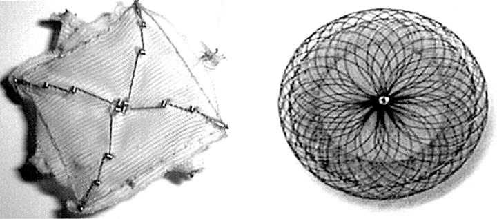
Figure 1 .
Atrial septal occluders. The Cardioseal occluder (left) is a spring loaded Dacron double umbrella. The Amplatzer occluder (right) is a circular double disk frame with a conjoint waist made of nitinol windings.
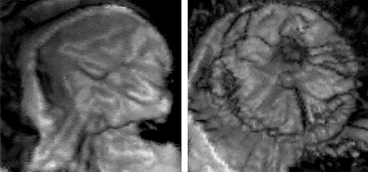
Figure 2 .
En face three dimensional views from the left atrium. The disk of the Cardioseal occluder (left) looks like a square and the four arms are applied to the left atrial side of the septum. The disk of the Amplatzer occluder (right) has a ball shape in the left atrial cavity.
Selected References
These references are in PubMed. This may not be the complete list of references from this article.
- Acar P., Laskari C., Rhodes J., Pandian N., Warner K., Marx G. Three-dimensional echocardiographic analysis of valve anatomy as a determinant of mitral regurgitation after surgery for atrioventricular septal defects. Am J Cardiol. 1999 Mar 1;83(5):745–749. doi: 10.1016/s0002-9149(98)00982-5. [DOI] [PubMed] [Google Scholar]
- Acar P., Maunoury C., Antonietti T., Bonnet D., Sidi D., Kachaner J. Left ventricular ejection fraction in children measured by three-dimensional echocardiography using a new transthoracic integrated 3D-probe. A comparison with equilibrium radionuclide angiography. Eur Heart J. 1998 Oct;19(10):1583–1588. doi: 10.1053/euhj.1998.1091. [DOI] [PubMed] [Google Scholar]
- Acar P., Saliba Z., Bonhoeffer P., Aggoun Y., Bonnet D., Sidi D., Kachaner J. Influence of atrial septal defect anatomy in patient selection and assessment of closure with the Cardioseal device; a three-dimensional transoesophageal echocardiographic reconstruction. Eur Heart J. 2000 Apr;21(7):573–581. doi: 10.1053/euhj.1999.1855. [DOI] [PubMed] [Google Scholar]
- Berger F., Ewert P., Björnstad P. G., Dähnert I., Krings G., Brilla-Austenat I., Vogel M., Lange P. E. Transcatheter closure as standard treatment for most interatrial defects: experience in 200 patients treated with the Amplatzer Septal Occluder. Cardiol Young. 1999 Sep;9(5):468–473. doi: 10.1017/s1047951100005369. [DOI] [PubMed] [Google Scholar]
- Chan K. C., Godman M. J., Walsh K., Wilson N., Redington A., Gibbs J. L. Transcatheter closure of atrial septal defect and interatrial communications with a new self expanding nitinol double disc device (Amplatzer septal occluder): multicentre UK experience. Heart. 1999 Sep;82(3):300–306. doi: 10.1136/hrt.82.3.300. [DOI] [PMC free article] [PubMed] [Google Scholar]
- Dall'Agata A., McGhie J., Taams M. A., Cromme-Dijkhuis A. H., Spitaels S. E., Breburda C. S., Roelandt J. R., Bogers A. J. Secundum atrial septal defect is a dynamic three-dimensional entity. Am Heart J. 1999 Jun;137(6):1075–1081. doi: 10.1016/s0002-8703(99)70365-0. [DOI] [PubMed] [Google Scholar]
- Dhillon R., Thanopoulos B., Tsaousis G., Triposkiadis F., Kyriakidis M., Redington A. Transcatheter closure of atrial septal defects in adults with the Amplatzer septal occluder. Heart. 1999 Nov;82(5):559–562. doi: 10.1136/hrt.82.5.559. [DOI] [PMC free article] [PubMed] [Google Scholar]
- Fischer G., Kramer H. H., Stieh J., Harding P., Jung O. Transcatheter closure of secundum atrial septal defects with the new self-centering Amplatzer Septal Occluder. Eur Heart J. 1999 Apr;20(7):541–549. doi: 10.1053/euhj.1998.1330. [DOI] [PubMed] [Google Scholar]
- Franke A., Kühl H. P., Rulands D., Jansen C., Erena C., Grabitz R. G., Däbritz S., Messmer B. J., Flachskampf F. A., Hanrath P. Quantitative analysis of the morphology of secundum-type atrial septal defects and their dynamic change using transesophageal three-dimensional echocardiography. Circulation. 1997 Nov 4;96(9 Suppl):II–323-7. [PubMed] [Google Scholar]
- Hausdorf G., Kaulitz R., Paul T., Carminati M., Lock J. Transcatheter closure of atrial septal defect with a new flexible, self-centering device (the STARFlex Occluder). Am J Cardiol. 1999 Nov 1;84(9):1113-6, A10. doi: 10.1016/s0002-9149(99)00515-9. [DOI] [PubMed] [Google Scholar]
- Kaulitz R., Paul T., Hausdorf G. Extending the limits of transcatheter closure of atrial septal defects with the double umbrella device (CardioSEAL). Heart. 1998 Jul;80(1):54–59. [PMC free article] [PubMed] [Google Scholar]
- Maeno Y. V., Benson L. N., Boutin C. Impact of dynamic 3D transoesophageal echocardiography in the assessment of atrial septal defects and occlusion by the double-umbrella device (CardioSEAL) Cardiol Young. 1998 Jul;8(3):368–378. doi: 10.1017/s1047951100006892. [DOI] [PubMed] [Google Scholar]
- Magni G., Hijazi Z. M., Pandian N. G., Delabays A., Sugeng L., Laskari C., Marx G. R. Two- and three-dimensional transesophageal echocardiography in patient selection and assessment of atrial septal defect closure by the new DAS-Angel Wings device: initial clinical experience. Circulation. 1997 Sep 16;96(6):1722–1728. doi: 10.1161/01.cir.96.6.1722. [DOI] [PubMed] [Google Scholar]
- Marx G. R., Fulton D. R., Pandian N. G., Vogel M., Cao Q. L., Ludomirsky A., Delabays A., Sugeng L., Klas B. Delineation of site, relative size and dynamic geometry of atrial septal defects by real-time three-dimensional echocardiography. J Am Coll Cardiol. 1995 Feb;25(2):482–490. doi: 10.1016/0735-1097(94)00372-w. [DOI] [PubMed] [Google Scholar]


