Abstract
OBJECTIVE—To use intravascular ultrasound (IVUS) to compare plaque morphology in acute myocardial infarction and stable angina pectoris. DESIGN—Retrospective study. SETTING—Primary care hospital. PATIENTS—59 consecutive cases of acute myocardial infarction and 50 consecutive cases of stable angina pectoris. METHODS—IVUS was used before coronary intervention. MAIN OUTCOME MEASURES—Plaque morphology (incidence of eccentric plaque, subtle dissections, low echoic thrombus, calcification, echolucent areas, and bright speckled echo material), assessed visually using IVUS. RESULTS—There were no significant differences in plaque eccentricity or calcification between the two groups, but low echoic thrombus (acute myocardial infarction 15% v stable angina pectoris 0%), subtle dissections (37% v 4%), echolucent areas (31% v 0%), and bright speckled echo material (90% v 0%) were more common in the infarction group than in the stable angina group (p < 0.001 for all). There was a longer time between the onset of symptoms and the IVUS examination in patients with low echoic thrombus than in those without (p < 0.03). CONCLUSIONS—Low echoic thrombus, subtle dissections, echolucent areas, and bright speckled echo material are morphological characteristics associated with plaque at the time of acute myocardial infarction. These findings correspond pathologically to ruptured plaque. Keywords: intravascular ultrasound; acute myocardial infarction; plaque morphology
Full Text
The Full Text of this article is available as a PDF (198.4 KB).
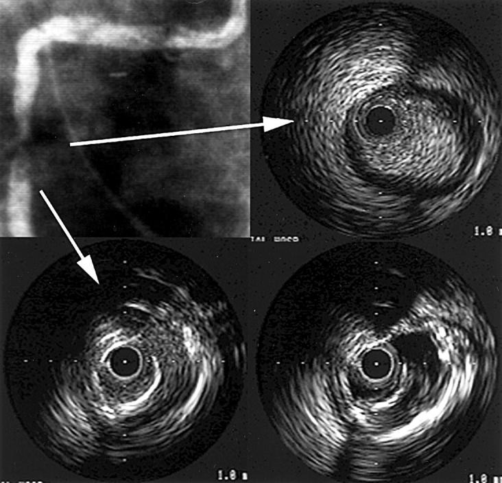
Figure 1 .

Coronary angiography and intravascular ultrasound (IVUS) images—1. The coronary angiography and IVUS images of a patient with acute myocardial infarction in the right coronary artery territory. (A, top left) Coronary angiography shows the right coronary artery with 99% stenosis in the mid-portion. (B, top right) A plain IVUS image of proximal stenosis. This shows bright speckled echo material. (C, bottom left) A plain IVUS image with suspected plaque dissection. (D, bottom right) Negative contrast image of (C). This reveals the plaque dissection at 3 o'clock more clearly than the plain image.
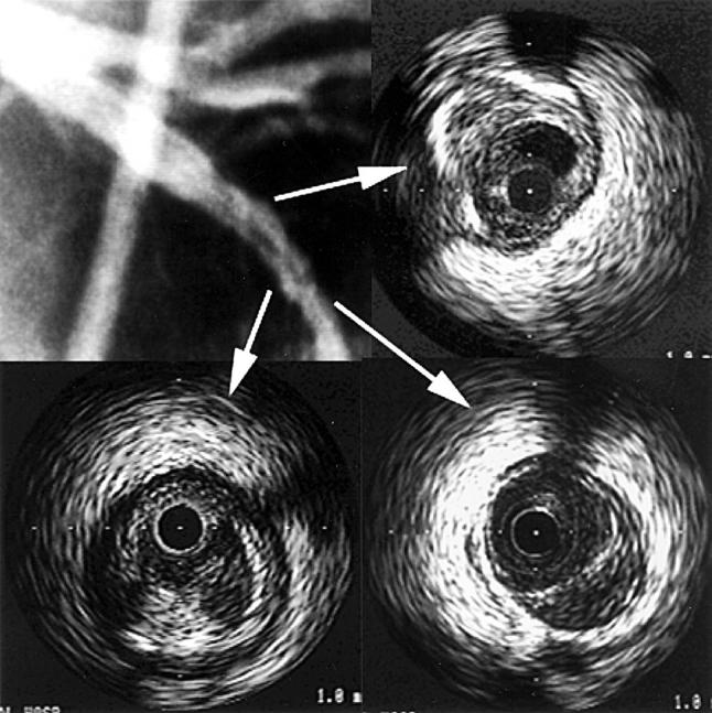
Figure 2 .

Coronary angiography and intravascular ultrasound (IVUS) images—2. The coronary angiography and IVUS images of a patient with acute myocardial infarction in left anterior descending coronary artery (LAD) territory. (A, top left) Coronary angiography shows filling defect in the mid-portion of the LAD. (B, top right) IVUS image of low echoic thrombus. In concordance with the angiographic findings, a low echoic mass representing thrombus can be seen at 12 to 2 o'clock. (C, bottom left) IVUS image with subtle dissection at 6 o'clock. (D, bottom right) IVUS image representing echolucent area at 4 to 6 o'clock; a low echoic lesion can be seen in the eccentric plaque.
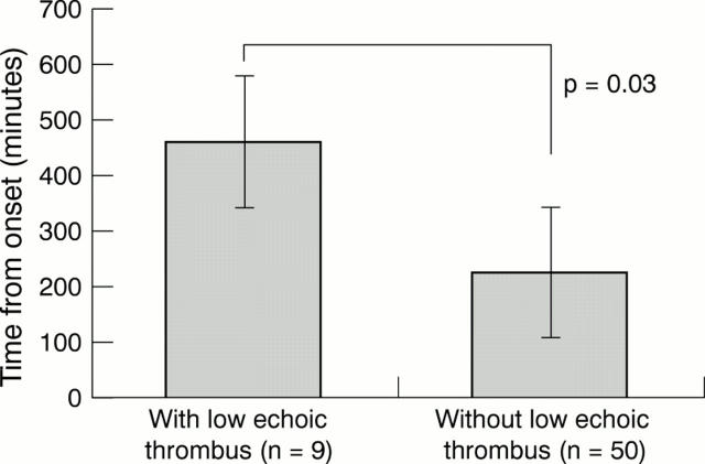
Figure 3 .
Comparison of time from onset of symptoms between cases with and without low echoic thrombus. The time course in cases with low echoic thrombus was significantly longer than in those without low echoic thrombus.
Selected References
These references are in PubMed. This may not be the complete list of references from this article.
- Abizaid A. S., Mintz G. S., Mehran R., Abizaid A., Lansky A. J., Pichard A. D., Satler L. F., Wu H., Pappas C., Kent K. M. Long-term follow-up after percutaneous transluminal coronary angioplasty was not performed based on intravascular ultrasound findings: importance of lumen dimensions. Circulation. 1999 Jul 20;100(3):256–261. doi: 10.1161/01.cir.100.3.256. [DOI] [PubMed] [Google Scholar]
- Albiero R., Rau T., Schlüter M., Di Mario C., Reimers B., Mathey D. G., Tobis J. M., Schofer J., Colombo A. Comparison of immediate and intermediate-term results of intravascular ultrasound versus angiography-guided Palmaz-Schatz stent implantation in matched lesions. Circulation. 1997 Nov 4;96(9):2997–3005. doi: 10.1161/01.cir.96.9.2997. [DOI] [PubMed] [Google Scholar]
- Alfonso F., Macaya C., Goicolea J., Iñiguez A., Hernandez R., Zamorano J., Perez-Vizcayne M. J., Zarco P. Intravascular ultrasound imaging of angiographically normal coronary segments in patients with coronary artery disease. Am Heart J. 1994 Mar;127(3):536–544. doi: 10.1016/0002-8703(94)90660-2. [DOI] [PubMed] [Google Scholar]
- Bhandari A. K., Nanda N. C., Hicks D. G. Two-dimensional echocardiography of intracardiac masses: echo pattern-histopathology correlation. Ultrasound Med Biol. 1982;8(6):673–680. doi: 10.1016/0301-5629(82)90124-7. [DOI] [PubMed] [Google Scholar]
- Bocksch W., Schartl M., Beckmann S., Dreysse S., Fleck E. Intravascular ultrasound assessment of direct percutaneous transluminal coronary angioplasty in patients with acute myocardial infarction. Coron Artery Dis. 1997 May;8(5):265–273. doi: 10.1097/00019501-199705000-00003. [DOI] [PubMed] [Google Scholar]
- Bocksch W., Schartl M., Beckmann S., Dreysse S., Fleck E. Intravascular ultrasound imaging in patients with acute myocardial infarction. Eur Heart J. 1995 Oct;16 (Suppl J):46–52. doi: 10.1093/eurheartj/16.suppl_j.46. [DOI] [PubMed] [Google Scholar]
- Brosius F. C., 3rd, Roberts W. C. Significance of coronary arterial thrombus in transmural acute myocardial infarction. A study of 54 necropsy patients. Circulation. 1981 Apr;63(4):810–816. doi: 10.1161/01.cir.63.4.810. [DOI] [PubMed] [Google Scholar]
- Bruchhäuser J., Sechtem U., Höpp H. W., Erdmann E. Intrakoronarer Ultraschall ändert therapeutische Strategie bei angiographisch mehrdeutigem Befund. Z Kardiol. 1997 Feb;86(2):138–147. doi: 10.1007/s003920050044. [DOI] [PubMed] [Google Scholar]
- Dangas G., Mintz G. S., Mehran R., Lansky A. J., Kornowski R., Pichard A. D., Satler L. F., Kent K. M., Stone G. W., Leon M. B. Preintervention arterial remodeling as an independent predictor of target-lesion revascularization after nonstent coronary intervention: an analysis of 777 lesions with intravascular ultrasound imaging. Circulation. 1999 Jun 22;99(24):3149–3154. doi: 10.1161/01.cir.99.24.3149. [DOI] [PubMed] [Google Scholar]
- De Scheerder I., De Man F., Herregods M. C., Wilczek K., Barrios L., Raymenants E., Desmet W., De Geest H., Piessens J. Intravascular ultrasound versus angiography for measurement of luminal diameters in normal and diseased coronary arteries. Am Heart J. 1994 Feb;127(2):243–251. doi: 10.1016/0002-8703(94)90110-4. [DOI] [PubMed] [Google Scholar]
- Erbel R., Ge J., Görge G., Baumgart D., Haude M., Jeremias A., von Birgelen C., Jollet N., Schwedtmann J. Intravascular ultrasound classification of atherosclerotic lesions according to American Heart Association recommendation. Coron Artery Dis. 1999 Oct;10(7):489–499. doi: 10.1097/00019501-199910000-00009. [DOI] [PubMed] [Google Scholar]
- Frimerman A., Miller H. I., Hallman M., Laniado S., Keren G. Intravascular ultrasound characterization of thrombi of different composition. Am J Cardiol. 1994 Jun 1;73(15):1053–1057. doi: 10.1016/0002-9149(94)90282-8. [DOI] [PubMed] [Google Scholar]
- Ge J., Baumgart D., Haude M., Görge G., von Birgelen C., Sack S., Erbel R. Role of intravascular ultrasound imaging in identifying vulnerable plaques. Herz. 1999 Feb;24(1):32–41. doi: 10.1007/BF03043816. [DOI] [PubMed] [Google Scholar]
- Ge J., Chirillo F., Schwedtmann J., Görge G., Haude M., Baumgart D., Shah V., von Birgelen C., Sack S., Boudoulas H. Screening of ruptured plaques in patients with coronary artery disease by intravascular ultrasound. Heart. 1999 Jun;81(6):621–627. doi: 10.1136/hrt.81.6.621. [DOI] [PMC free article] [PubMed] [Google Scholar]
- Ge J., Erbel R., Gerber T., Görge G., Koch L., Haude M., Meyer J. Intravascular ultrasound imaging of angiographically normal coronary arteries: a prospective study in vivo. Br Heart J. 1994 Jun;71(6):572–578. doi: 10.1136/hrt.71.6.572. [DOI] [PMC free article] [PubMed] [Google Scholar]
- Görge G., Ge J., Haude M., Shah V., Jeremias A., Simon H., Erbel R. Intravascular ultrasound for evaluation of coronary arteries. Herz. 1996 Apr;21(2):78–89. [PubMed] [Google Scholar]
- Hoffmann R., Mintz G. S., Mehran R., Kent K. M., Pichard A. D., Satler L. F., Leon M. B. Tissue proliferation within and surrounding Palmaz-Schatz stents is dependent on the aggressiveness of stent implantation technique. Am J Cardiol. 1999 Apr 15;83(8):1170–1174. doi: 10.1016/s0002-9149(99)00053-3. [DOI] [PubMed] [Google Scholar]
- Honye J., Saito S., Takayama T., Yajima J., Shimizu T., Chiku M., Mizumura T., Takaiwa Y., Horiuchi K., Moriuchi M. Clinical utility of negative contrast intravascular ultrasound to evaluate plaque morphology before and after coronary interventions. Am J Cardiol. 1999 Mar 1;83(5):687–690. doi: 10.1016/s0002-9149(98)00971-0. [DOI] [PubMed] [Google Scholar]
- Kojima S., Nonogi H., Miyao Y., Miyazaki S., Goto Y., Itoh A., Daikoku S., Matsumoto T., Morii I., Yutani C. Is preinfarction angina related to the presence or absence of coronary plaque rupture? Heart. 2000 Jan;83(1):64–68. doi: 10.1136/heart.83.1.64. [DOI] [PMC free article] [PubMed] [Google Scholar]
- Metz J. A., Yock P. G., Fitzgerald P. J. Intravascular ultrasound: basic interpretation. Cardiol Clin. 1997 Feb;15(1):1–15. doi: 10.1016/s0733-8651(05)70314-3. [DOI] [PubMed] [Google Scholar]
- Namiki N., Uchiyama T., Nagai Y., Yamashina A. Graphical comparison of coronary arterial culprit lesions in acute myocardial infarction and unstable angina pectoris. Intern Med. 1999 Nov;38(11):849–855. doi: 10.2169/internalmedicine.38.849. [DOI] [PubMed] [Google Scholar]
- Schroeder S., Baumbach A., Haase K. K., Oberhoff M., Marholdt H., Herdeg C., Athanasiadis A., Karsch K. R. Reduction of restenosis by vessel size adapted percutaneous transluminal coronary angioplasty using intravascular ultrasound. Am J Cardiol. 1999 Mar 15;83(6):875–879. doi: 10.1016/s0002-9149(98)01069-8. [DOI] [PubMed] [Google Scholar]
- Weissman N. J., Sheris S. J., Chari R., Mendelsohn F. O., Anderson W. D., Breall J. A., Tanguay J. F., Diver D. J. Intravascular ultrasonic analysis of plaque characteristics associated with coronary artery remodeling. Am J Cardiol. 1999 Jul 1;84(1):37–40. doi: 10.1016/s0002-9149(99)00188-5. [DOI] [PubMed] [Google Scholar]
- Yamagishi M., Terashima M., Awano K., Kijima M., Nakatani S., Daikoku S., Ito K., Yasumura Y., Miyatake K. Morphology of vulnerable coronary plaque: insights from follow-up of patients examined by intravascular ultrasound before an acute coronary syndrome. J Am Coll Cardiol. 2000 Jan;35(1):106–111. doi: 10.1016/s0735-1097(99)00533-1. [DOI] [PubMed] [Google Scholar]
- Yock P. G. The future of minimally invasive myocardial revascularization: a cardiologist's view. J Card Surg. 1998 Jul;13(4):310–315. doi: 10.1111/j.1540-8191.1998.tb01075.x. [DOI] [PubMed] [Google Scholar]
- van der Wal A. C., Becker A. E., van der Loos C. M., Das P. K. Site of intimal rupture or erosion of thrombosed coronary atherosclerotic plaques is characterized by an inflammatory process irrespective of the dominant plaque morphology. Circulation. 1994 Jan;89(1):36–44. doi: 10.1161/01.cir.89.1.36. [DOI] [PubMed] [Google Scholar]


