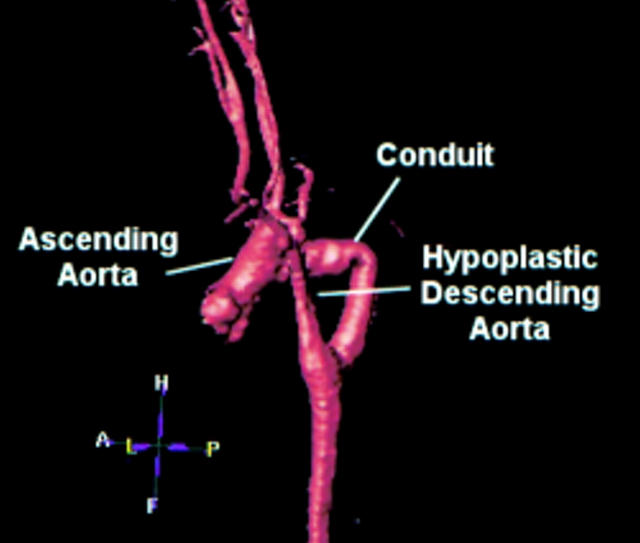
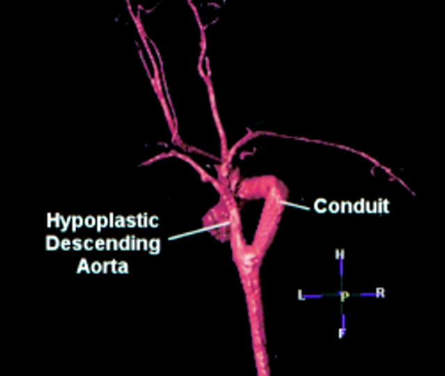
(Left) Left lateral view of three dimensional reconstructed gadolinium enhanced magnetic resonance angiogram showing the right aortic arch and the hypoplastic descending aorta. Note the residual coarctation between the right common carotid and subclavian arteries, and the additional areas of stenosis in the right subclavian artery and descending aorta. The mid-conduit stenosis can be clearly seen. There is a retro-oesophageal left subclavian artery.(Right) Posterior view showing the residual coarctation and the mid-conduit stenosis. A, anterior; P, posterior; L, left, R, right, H, head, F, foot.

An official website of the United States government
Here's how you know
Official websites use .gov
A
.gov website belongs to an official
government organization in the United States.
Secure .gov websites use HTTPS
A lock (
) or https:// means you've safely
connected to the .gov website. Share sensitive
information only on official, secure websites.

