Abstract
OBJECTIVE—To evaluate new echocardiographic modes in the diagnosis of arrhythmogenic right ventricular cardiomyopathy (ARVC). DESIGN—Prospective observational study. SETTING—University Hospital. SUBJECTS—15 patients with ARVC and a control group of 25 healthy subjects. METHODS—Transthoracic echocardiography included cross sectional measurements of the right ventricular outflow tract, right ventricular inflow tract, and right ventricular body. Wall motion was analysed subjectively. M mode and pulsed tissue Doppler techniques were used for quantitative measurement of tricuspid annular motion at the lateral, septal, posterior, and anterior positions. Doppler assessment of tricuspid flow and systemic venous flow was also performed. RESULTS—Assessed by M mode, the total amplitude of the tricuspid annular motion was significantly decreased in the lateral, septal, and posterior positions in the patients compared with the controls. The tissue Doppler velocity pattern showed decreased early diastolic peak annular (EA) velocity and an accompanying decrease in early (EA) to late diastolic (AA) velocity ratio in all positions; the systolic annular velocity was significantly decreased only in the lateral position. Four patients had normal right ventricular dimensions and three were judged to have normal right ventricular wall motion. The patient group had also a significantly decreased tricuspid flow E:A ratio. CONCLUSIONS—Tricuspid annular measurements are valuable, easy to obtain, and allow quantitative assessment of right ventricular function. ARVC patients showed an abnormal velocity pattern that may be an early but non-specific sign of the disease. Normal right ventricular dimensions do not exclude ARVC, and subjective detection of early changes in wall motion may be difficult. Keywords: annular motion; diastolic dysfunction; right ventricular function; tissue Doppler
Full Text
The Full Text of this article is available as a PDF (192.3 KB).
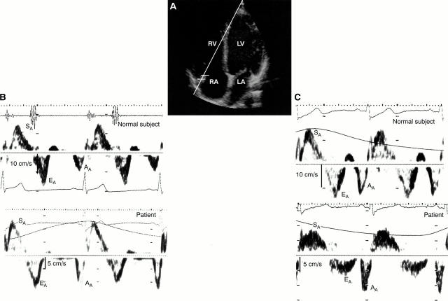
Figure 1 .
(A) Pulsed tissue Doppler recording of lateral tricuspid annulus motion. Guided by the cross sectional apical image, the sample volume was placed in the bright area of the tricuspid annulus, trying to align the cursor as parallel to the motion as possible. (B) Pulsed tissue Doppler recordings from a normal subject aged 26 years and a patient aged 27 years. (C) Pulsed tissue Doppler recordings from a normal subject aged 47 years and a patient aged 49 years. Note the decreased early diastolic annular velocity with increasing age in normal subjects and in patients with arrhythmogenic right ventricular cardiomyopathy compared with age matched normal controls. The systolic peak annular velocity is also slightly decreased in the patients. AA, late diastolic peak annular velocity; EA, early diastolic peak annular velocity; LA, left atrium; LV, left ventricle; RA, right atrium; RV, right ventricle; SA, systolic peak annular velocity.
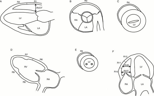
Figure 2 .
Definition of wall segments and cavity dimensions of the right ventricle. (A) Parasternal long axis view of the left heart: right ventricular outflow tract, measured from the anterior right ventricular free wall to the right side of the interventricular septum. (B) Parasternal short axis view at the aortic root level. (C) Parasternal short axis view at the mitral valve level. (D) Parasternal long axis view of the right ventricular inflow tract. (E) Parasternal short axis view at the papillary muscle level. (F) Apical four chamber view: measurements of right ventricular inflow tract were taken within one third of the distance below the tricuspid annulus towards the right ventricular apex, and of the right ventricular body from the maximum short axis dimension in the middle third of the right ventricle. Segments R1, R2, and R3 correspond to the anterior wall of the outflow tract, R4, R6, and R7 to the anterior free wall, R5, R8, and R9 to the inferior wall, and R10, R11, and R12 to the lateral free wall of the right ventricle.13 LA, left atrium; LV, left ventricle; R, right ventricular segment; RA, right atrium; RV, right ventricle; RVB, right ventricular body; RVIT, right ventricular inflow tract; RVOT, right ventricular outflow tract.
Selected References
These references are in PubMed. This may not be the complete list of references from this article.
- Alam M., Wardell J., Andersson E., Samad B. A., Nordlander R. Characteristics of mitral and tricuspid annular velocities determined by pulsed wave Doppler tissue imaging in healthy subjects. J Am Soc Echocardiogr. 1999 Aug;12(8):618–628. doi: 10.1053/je.1999.v12.a99246. [DOI] [PubMed] [Google Scholar]
- Appleton C. P., Jensen J. L., Hatle L. K., Oh J. K. Doppler evaluation of left and right ventricular diastolic function: a technical guide for obtaining optimal flow velocity recordings. J Am Soc Echocardiogr. 1997 Apr;10(3):271–292. doi: 10.1016/s0894-7317(97)70063-4. [DOI] [PubMed] [Google Scholar]
- Blomström-Lundqvist C., Beckman-Suurküla M., Wallentin I., Jonsson R., Olsson S. B. Ventricular dimensions and wall motion assessed by echocardiography in patients with arrhythmogenic right ventricular dysplasia. Eur Heart J. 1988 Dec;9(12):1291–1302. doi: 10.1093/oxfordjournals.eurheartj.a062446. [DOI] [PubMed] [Google Scholar]
- Foale R., Nihoyannopoulos P., McKenna W., Kleinebenne A., Nadazdin A., Rowland E., Smith G., Klienebenne A. Echocardiographic measurement of the normal adult right ventricle. Br Heart J. 1986 Jul;56(1):33–44. doi: 10.1136/hrt.56.1.33. [DOI] [PMC free article] [PubMed] [Google Scholar]
- Furlanello F., Bertoldi A., Dallago M., Furlanello C., Fernando F., Inama G., Pappone C., Chierchia S. Cardiac arrest and sudden death in competitive athletes with arrhythmogenic right ventricular dysplasia. Pacing Clin Electrophysiol. 1998 Jan;21(1 Pt 2):331–335. doi: 10.1111/j.1540-8159.1998.tb01116.x. [DOI] [PubMed] [Google Scholar]
- Goldberger J. J., Himelman R. B., Wolfe C. L., Schiller N. B. Right ventricular infarction: recognition and assessment of its hemodynamic significance by two-dimensional echocardiography. J Am Soc Echocardiogr. 1991 Mar-Apr;4(2):140–146. doi: 10.1016/s0894-7317(14)80525-7. [DOI] [PubMed] [Google Scholar]
- Gulati V. K., Katz W. E., Follansbee W. P., Gorcsan J., 3rd Mitral annular descent velocity by tissue Doppler echocardiography as an index of global left ventricular function. Am J Cardiol. 1996 May 1;77(11):979–984. doi: 10.1016/s0002-9149(96)00033-1. [DOI] [PubMed] [Google Scholar]
- Hammarström E., Wranne B., Pinto F. J., Puryear J., Popp R. L. Tricuspid annular motion. J Am Soc Echocardiogr. 1991 Mar-Apr;4(2):131–139. doi: 10.1016/s0894-7317(14)80524-5. [DOI] [PubMed] [Google Scholar]
- Iliceto S., Izzi M., De Martino G., Rizzon P. Echo Doppler evaluation of right ventricular dysplasia. Eur Heart J. 1989 Sep;10 (Suppl 500):29–32. doi: 10.1093/eurheartj/10.suppl_d.29. [DOI] [PubMed] [Google Scholar]
- Isaaz K., Munoz del Romeral L., Lee E., Schiller N. B. Quantitation of the motion of the cardiac base in normal subjects by Doppler echocardiography. J Am Soc Echocardiogr. 1993 Mar-Apr;6(2):166–176. doi: 10.1016/s0894-7317(14)80487-2. [DOI] [PubMed] [Google Scholar]
- Kaul S., Tei C., Hopkins J. M., Shah P. M. Assessment of right ventricular function using two-dimensional echocardiography. Am Heart J. 1984 Mar;107(3):526–531. doi: 10.1016/0002-8703(84)90095-4. [DOI] [PubMed] [Google Scholar]
- Kukulski T., Hübbert L., Arnold M., Wranne B., Hatle L., Sutherland G. R. Normal regional right ventricular function and its change with age: a Doppler myocardial imaging study. J Am Soc Echocardiogr. 2000 Mar;13(3):194–204. doi: 10.1067/mje.2000.103106. [DOI] [PubMed] [Google Scholar]
- Larsson E., Wesslén L., Lindquist O., Baandrup U., Eriksson L., Olsen E., Rolf C., Friman G. Sudden unexpected cardiac deaths among young Swedish orienteers--morphological changes in hearts and other organs. APMIS. 1999 Mar;107(3):325–336. doi: 10.1111/j.1699-0463.1999.tb01561.x. [DOI] [PubMed] [Google Scholar]
- Lindström L., Wranne B. Pulsed tissue Doppler evaluation of mitral annulus motion: a new window to assessment of diastolic function. Clin Physiol. 1999 Jan;19(1):1–10. doi: 10.1046/j.1365-2281.1999.00137.x. [DOI] [PubMed] [Google Scholar]
- Manyari D. E., Duff H. J., Kostuk W. J., Belenkie I., Klein G. J., Wyse D. G., Mitchell L. B., Boughner D., Guiraudon G., Smith E. R. Usefulness of noninvasive studies for diagnosis of right ventricular dysplasia. Am J Cardiol. 1986 May 1;57(13):1147–1153. doi: 10.1016/0002-9149(86)90690-9. [DOI] [PubMed] [Google Scholar]
- Marcus F. I., Fontaine G. H., Guiraudon G., Frank R., Laurenceau J. L., Malergue C., Grosgogeat Y. Right ventricular dysplasia: a report of 24 adult cases. Circulation. 1982 Feb;65(2):384–398. doi: 10.1161/01.cir.65.2.384. [DOI] [PubMed] [Google Scholar]
- McKenna W. J., Thiene G., Nava A., Fontaliran F., Blomstrom-Lundqvist C., Fontaine G., Camerini F. Diagnosis of arrhythmogenic right ventricular dysplasia/cardiomyopathy. Task Force of the Working Group Myocardial and Pericardial Disease of the European Society of Cardiology and of the Scientific Council on Cardiomyopathies of the International Society and Federation of Cardiology. Br Heart J. 1994 Mar;71(3):215–218. doi: 10.1136/hrt.71.3.215. [DOI] [PMC free article] [PubMed] [Google Scholar]
- Mendes L. A., Dec G. W., Picard M. H., Palacios I. F., Newell J., Davidoff R. Right ventricular dysfunction: an independent predictor of adverse outcome in patients with myocarditis. Am Heart J. 1994 Aug;128(2):301–307. doi: 10.1016/0002-8703(94)90483-9. [DOI] [PubMed] [Google Scholar]
- Pinamonti B., Di Lenarda A., Sinagra G., Silvestri F., Bussani R., Camerini F. Long-term evolution of right ventricular dysplasia-cardiomyopathy. The Heart Muscle Disease Study Group. Am Heart J. 1995 Feb;129(2):412–415. doi: 10.1016/0002-8703(95)90029-2. [DOI] [PubMed] [Google Scholar]
- Richardson P., McKenna W., Bristow M., Maisch B., Mautner B., O'Connell J., Olsen E., Thiene G., Goodwin J., Gyarfas I. Report of the 1995 World Health Organization/International Society and Federation of Cardiology Task Force on the Definition and Classification of cardiomyopathies. Circulation. 1996 Mar 1;93(5):841–842. doi: 10.1161/01.cir.93.5.841. [DOI] [PubMed] [Google Scholar]
- Scognamiglio R., Fasoli G., Nava A., Miraglia G., Thiene G., Dalla-Volta S. Contribution of cross-sectional echocardiography to the diagnosis of right ventricular dysplasia at the asymptomatic stage. Eur Heart J. 1989 Jun;10(6):538–542. doi: 10.1093/oxfordjournals.eurheartj.a059524. [DOI] [PubMed] [Google Scholar]
- Sivaciyan V., Ranganathan N. Transcutaneous doppler jugular venous flow velocity recording. Circulation. 1978 May;57(5):930–939. doi: 10.1161/01.cir.57.5.930. [DOI] [PubMed] [Google Scholar]
- Thiene G., Nava A., Corrado D., Rossi L., Pennelli N. Right ventricular cardiomyopathy and sudden death in young people. N Engl J Med. 1988 Jan 21;318(3):129–133. doi: 10.1056/NEJM198801213180301. [DOI] [PubMed] [Google Scholar]
- Wilkenshoff U. M., Sovany A., Wigström L., Olstad B., Lindström L., Engvall J., Janerot-Sjöberg B., Wranne B., Hatle L., Sutherland G. R. Regional mean systolic myocardial velocity estimation by real-time color Doppler myocardial imaging: a new technique for quantifying regional systolic function. J Am Soc Echocardiogr. 1998 Jul;11(7):683–692. doi: 10.1053/je.1998.v11.a90584. [DOI] [PubMed] [Google Scholar]


