Abstract
BACKGROUND—Biventricular pacing has been proposed as an adjuvant to optimal medical treatment in patients with drug refractory heart failure caused by chronic left ventricular systolic dysfunction and intraventricular conduction delay. OBJECTIVE—To assess the technical feasibility and long term results (over six years) of transverse left ventricular pacing with the lead inserted into a tributary vein of the coronary sinus. SUBJECTS—From August 1994 to February 2000, left ventricular lead implantation was attempted in 116 patients who were eligible for biventricular pacing (mean (SD) age 67 (9) years, New York Heart Association (NYHA) functional class III/IV, left ventricular ejection fraction 22 (6)%, QRS duration 185 (26) ms). RESULTS—The overall implantation success rate was 88% (n = 102). A learning curve was indicated by a progressive increase in success from 61% early on to 98% in the last year. The mean pacing threshold was 1.1 (0.7) V/0.5 ms at the time of implantation and increased slightly up to 1.9 (0.9) V/0.5 ms at the end of the follow up period (15 (13) months). The rate of acute and delayed left ventricular lead dislodgement decreased from 30% in the early years to 11% after 1999. During follow up, 19 patients required reoperation for delayed lead dislodgement or increase in left ventricular pacing threshold (n = 15), phrenic nerve stimulation (n = 3), or infection (n = 3). CONCLUSIONS—Transverse left ventricular pacing through the coronary sinus is feasible and safe. The rate of implantation failure and of lead related problems has decreased greatly with increasing experience and with improvements in the equipment. Keywords: biventricular pacing; left ventricular pacing; heart failure; intraventricular conduction delay
Full Text
The Full Text of this article is available as a PDF (202.7 KB).
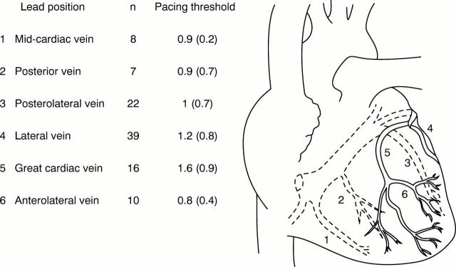
Figure 1 .
Left ventricular lead position and implant pacing thresholds.
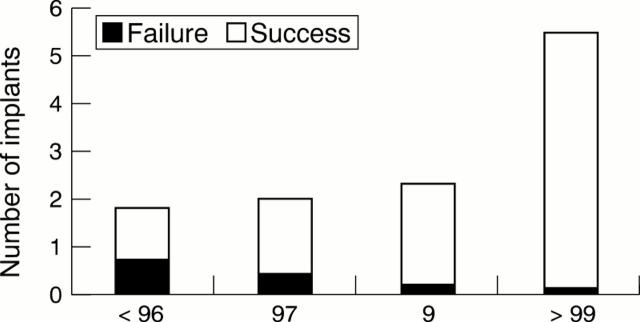
Figure 2 .
Change in implantation rate over time.
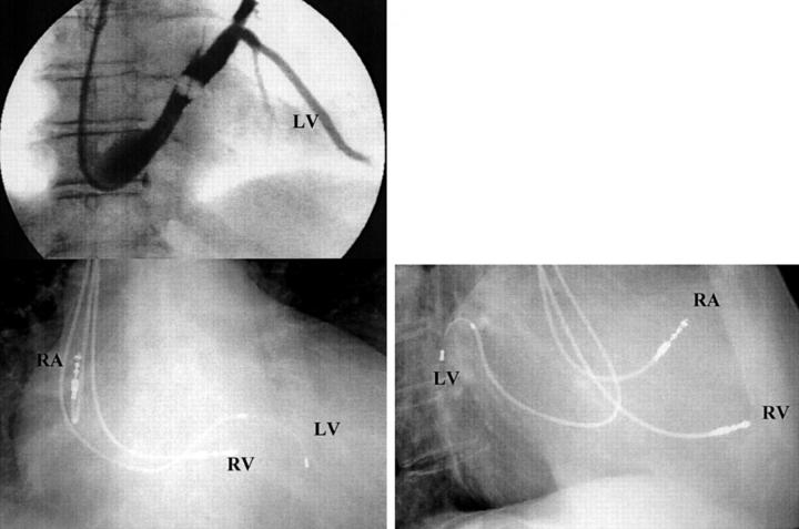
Figure 3 .
In this example there is a large lateral vein in which a thick conventional lead could easily be introduced. LV, lateral vein; RA, right atrial lead; RV, right ventricular lead; LV, left ventricular lead.
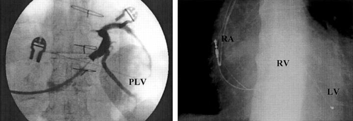
Figure 4 .
In this coronary sinus angiogram (left) there was a large posterolateral vein. However, the angle it makes with the coronary sinus may make it difficult to reach. In this case a very thin lead, with side wire technology, could be introduced into the vein (chest x ray on the right). LV, left ventricular lead; PLV, posterolateral vein; RA, right atrial lead; RV, right ventricular lead.
Figure 5 .
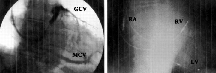
It can be seen on this coronary sinus angiogram (left) that there was no lateral or posterolateral vein, so that a choice had to be made between the great cardiac vein and the mid-cardiac vein. In this case the left ventricular lead was placed in the mid-cardiac vein, as can be seen on the chest x ray (right). GCV, great cardiac vein; LV, left ventricular lead; MCV, mid-cardiac vein; RA, right atrial lead; RV, right ventricular lead.
Selected References
These references are in PubMed. This may not be the complete list of references from this article.
- Auricchio A., Stellbrink C., Block M., Sack S., Vogt J., Bakker P., Klein H., Kramer A., Ding J., Salo R. Effect of pacing chamber and atrioventricular delay on acute systolic function of paced patients with congestive heart failure. The Pacing Therapies for Congestive Heart Failure Study Group. The Guidant Congestive Heart Failure Research Group. Circulation. 1999 Jun 15;99(23):2993–3001. doi: 10.1161/01.cir.99.23.2993. [DOI] [PubMed] [Google Scholar]
- Bakker P. F., Meijburg H. W., de Vries J. W., Mower M. M., Thomas A. C., Hull M. L., Robles De Medina E. O., Bredée J. J. Biventricular pacing in end-stage heart failure improves functional capacity and left ventricular function. J Interv Card Electrophysiol. 2000 Jun;4(2):395–404. doi: 10.1023/a:1009854417694. [DOI] [PubMed] [Google Scholar]
- Blanc J. J., Benditt D. G., Gilard M., Etienne Y., Mansourati J., Lurie K. G. A method for permanent transvenous left ventricular pacing. Pacing Clin Electrophysiol. 1998 Nov;21(11 Pt 1):2021–2024. doi: 10.1111/j.1540-8159.1998.tb01119.x. [DOI] [PubMed] [Google Scholar]
- Blanc J. J., Etienne Y., Gilard M., Mansourati J., Munier S., Boschat J., Benditt D. G., Lurie K. G. Evaluation of different ventricular pacing sites in patients with severe heart failure: results of an acute hemodynamic study. Circulation. 1997 Nov 18;96(10):3273–3277. doi: 10.1161/01.cir.96.10.3273. [DOI] [PubMed] [Google Scholar]
- Cazeau S., Ritter P., Bakdach S., Lazarus A., Limousin M., Henao L., Mundler O., Daubert J. C., Mugica J. Four chamber pacing in dilated cardiomyopathy. Pacing Clin Electrophysiol. 1994 Nov;17(11 Pt 2):1974–1979. doi: 10.1111/j.1540-8159.1994.tb03783.x. [DOI] [PubMed] [Google Scholar]
- Daubert J. C., Cazeau S., Leclercq C. Do we have reasons to be enthusiastic about pacing to treat advanced heart failure? Eur J Heart Fail. 1999 Aug;1(3):281–287. doi: 10.1016/s1388-9842(99)00042-2. [DOI] [PubMed] [Google Scholar]
- Daubert J. C., Ritter P., Le Breton H., Gras D., Leclercq C., Lazarus A., Mugica J., Mabo P., Cazeau S. Permanent left ventricular pacing with transvenous leads inserted into the coronary veins. Pacing Clin Electrophysiol. 1998 Jan;21(1 Pt 2):239–245. doi: 10.1111/j.1540-8159.1998.tb01096.x. [DOI] [PubMed] [Google Scholar]
- Gilard M., Mansourati J., Etienne Y., Larlet J. M., Truong B., Boschat J., Blanc J. J. Angiographic anatomy of the coronary sinus and its tributaries. Pacing Clin Electrophysiol. 1998 Nov;21(11 Pt 2):2280–2284. doi: 10.1111/j.1540-8159.1998.tb01167.x. [DOI] [PubMed] [Google Scholar]
- Gras D., Mabo P., Tang T., Luttikuis O., Chatoor R., Pedersen A. K., Tscheliessnigg H. H., Deharo J. C., Puglisi A., Silvestre J. Multisite pacing as a supplemental treatment of congestive heart failure: preliminary results of the Medtronic Inc. InSync Study. Pacing Clin Electrophysiol. 1998 Nov;21(11 Pt 2):2249–2255. doi: 10.1111/j.1540-8159.1998.tb01162.x. [DOI] [PubMed] [Google Scholar]
- Jaïs P., Douard H., Shah D. C., Barold S., Barat J. L., Clémenty J. Endocardial biventricular pacing. Pacing Clin Electrophysiol. 1998 Nov;21(11 Pt 1):2128–2131. doi: 10.1111/j.1540-8159.1998.tb01133.x. [DOI] [PubMed] [Google Scholar]
- Kass D. A., Chen C. H., Curry C., Talbot M., Berger R., Fetics B., Nevo E. Improved left ventricular mechanics from acute VDD pacing in patients with dilated cardiomyopathy and ventricular conduction delay. Circulation. 1999 Mar 30;99(12):1567–1573. doi: 10.1161/01.cir.99.12.1567. [DOI] [PubMed] [Google Scholar]
- Kindermann M., Fröhlig G., Doerr T., Schieffer H. Optimizing the AV delay in DDD pacemaker patients with high degree AV block: mitral valve Doppler versus impedance cardiography. Pacing Clin Electrophysiol. 1997 Oct;20(10 Pt 1):2453–2462. doi: 10.1111/j.1540-8159.1997.tb06085.x. [DOI] [PubMed] [Google Scholar]
- Kiviniemi M. S., Pirnes M. A., Eränen H. J., Kettunen R. V., Hartikainen J. E. Complications related to permanent pacemaker therapy. Pacing Clin Electrophysiol. 1999 May;22(5):711–720. doi: 10.1111/j.1540-8159.1999.tb00534.x. [DOI] [PubMed] [Google Scholar]
- Leclercq C., Cazeau S., Le Breton H., Ritter P., Mabo P., Gras D., Pavin D., Lazarus A., Daubert J. C. Acute hemodynamic effects of biventricular DDD pacing in patients with end-stage heart failure. J Am Coll Cardiol. 1998 Dec;32(7):1825–1831. doi: 10.1016/s0735-1097(98)00492-6. [DOI] [PubMed] [Google Scholar]
- Rosenbush S. W., Ruggie N., Turner D. A., Von Behren P. L., Denes P., Fordham E. W., Groch M. W., Messer J. V. Sequence and timing of ventricular wall motion in patients with bundle branch block. Assessment by radionuclide cineangiography. Circulation. 1982 Nov;66(5):1113–1119. doi: 10.1161/01.cir.66.5.1113. [DOI] [PubMed] [Google Scholar]
- Walker S., Levy T., Rex S., Paul V. E. The use of a "side-wire" permanent transvenous pacing electrode for left ventricular pacing. Europace. 1999 Jul;1(3):197–200. doi: 10.1053/eupc.1999.0039. [DOI] [PubMed] [Google Scholar]


