Abstract
A patient with cardiac arrest and documented torsade de pointes ventricular tachycardia is presented in whom acute coxsackievirus B2 myocarditis was identified as the most likely underlying cardiac condition. This case shows that torsade de pointes may occur as a rare manifestation of viral myocarditis. Serial serological tests and endomyocardial biopsies may be helpful in establishing a diagnosis in such patients. Keywords: torsade de pointes; ventricular tachycardia; viral myocarditis
Full Text
The Full Text of this article is available as a PDF (124.6 KB).
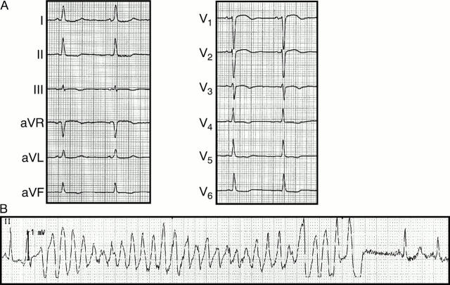
Figure 1 .
(A) Initial 12 lead ECG (25 mm/s) with anterior T wave inversions. (B) Torsade de pointes like ventricular tachycardia. The sinus beat following spontaneous termination of the tachycardia shows a prolonged QT duration.
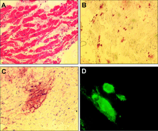
Figure 2 .
Endomyocardial biopsy results. (A) Haematoxylin and eosin staining, ×200. (B) CD45 immunostaining with horseradish peroxidase signal detection, ×200. Note the abundance and foci of inflammatory cells. (C) Immunostaining for the enteroviral VP-1 antigen with horseradish peroxidase signal detection, ×200. Note the strong sarcolemmal staining pattern in a group of cardiomyocytes. (D) VP-1 immunofluorescence staining of a mouse heart experimentally infected with coxsackievirus B3, ×400. The virally infected cells show a strong signal similar to the pattern described in C.


