Abstract
AIM—To determine clear cut echocardiographic criteria for isolated ventricular non-compaction (IVNC), a cardiomyopathy as yet "unclassified" by the World Health Organization. The disease is not widely known and its diagnosis mostly missed. METHODS AND RESULTS—In seven out of a series of 34 patients with IVNC the in vivo echocardiographic characteristics were validated against the anatomical examination of the heart removed after death in four and due to heart transplantation in three patients. Four morphological criteria diagnostic for IVNC were found. (1) Coexisting cardiac abnormalities were absent (by definition). (2) A two layer structure was seen, with a compacted thin epicardial band and a much thicker non-compacted endocardial layer of trabecular meshwork with deep endomyocardial spaces. A maximal end systolic ratio of non-compacted to compacted layers of > 2 is diagnostic. (3) The predominant localisation of the pathology was to mid-lateral (seven of seven patients), apical (six), and mid-inferior (seven) areas. The pathological preparations confirmed the echocardiographic findings. Concomitant regional hypokinesia was not confined to the non-compacted segments. (4) There was colour Doppler evidence of deep perfused intertrabecular recesses. CONCLUSIONS—Four clear cut echocardiographic diagnostic criteria were established. It is suggested that the WHO classification of cardiomyopathies be reconsidered to include IVNC as a distinct cardiomyopathy. Keywords: isolated ventricular non-compaction; morphological criteria; cardiomyopathy; echocardiography; pathology
Full Text
The Full Text of this article is available as a PDF (258.0 KB).
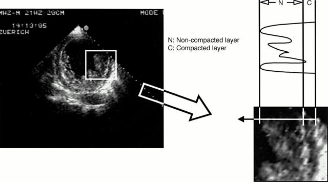
Figure 1 .
To quantify the extent of non-compaction at the site of maximal wall thickness the end systolic ratio of non-compacted to compacted thickness was determined. The two layers are best visualised at end systole as shown in this short axis view.
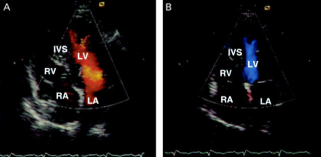
Figure 2 .
Colour Doppler study showed typical forward blood flow from the ventricular cavity into the deep spaces between the prominent trabeculation during diastole (in A represented by a red signal) with a reversed flow back into the ventricle during systole (in B, blue signal).
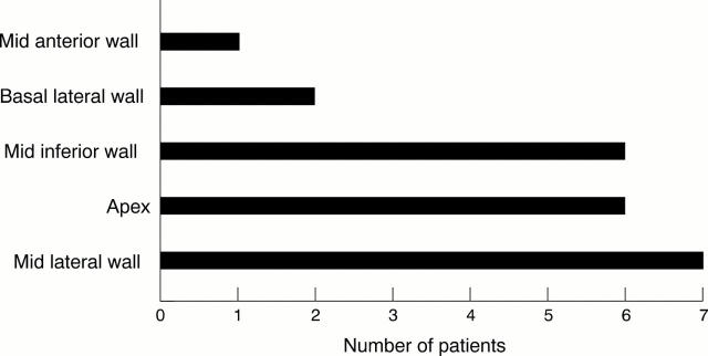
Figure 3 .
The structural alterations were predominantly localised to the left ventricular mid lateral wall and to the apex and the mid inferior wall.
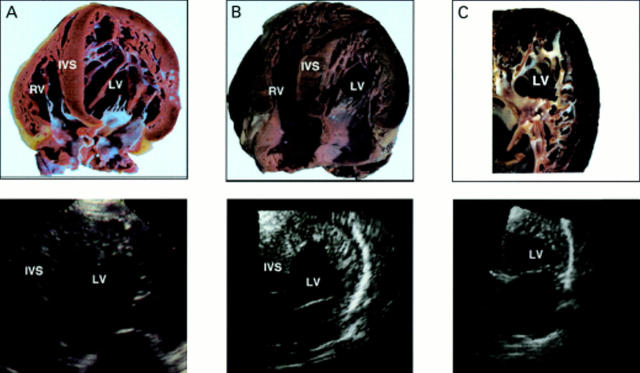
Figure 4 .
Apical four chamber view of three hearts. The anatomical findings (upper panel) are in agreement with the findings of the previously recorded echocardiographic view in the same patient (lower panel).
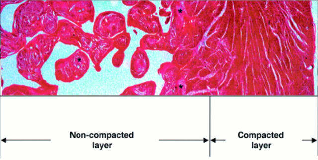
Figure 5 .
Histological preparation from the left ventricular apex of a patient with isolated ventricular non-compaction. Note the thin compacted normal outer layer of myocardium and the endocardial (non-compacted) layer. There is scar tissue within the trabeculations (asterisks) and in the subendocardial area but not in the epicardial zone.
Selected References
These references are in PubMed. This may not be the complete list of references from this article.
- Allenby P. A., Gould N. S., Schwartz M. F., Chiemmongkoltip P. Dysplastic cardiac development presenting as cardiomyopathy. Arch Pathol Lab Med. 1988 Dec;112(12):1255–1258. [PubMed] [Google Scholar]
- Angelini A., Melacini P., Barbero F., Thiene G. Evolutionary persistence of spongy myocardium in humans. Circulation. 1999 May 11;99(18):2475–2475. doi: 10.1161/01.cir.99.18.2475. [DOI] [PubMed] [Google Scholar]
- Boyd M. T., Seward J. B., Tajik A. J., Edwards W. D. Frequency and location of prominent left ventricular trabeculations at autopsy in 474 normal human hearts: implications for evaluation of mural thrombi by two-dimensional echocardiography. J Am Coll Cardiol. 1987 Feb;9(2):323–326. doi: 10.1016/s0735-1097(87)80383-2. [DOI] [PubMed] [Google Scholar]
- Chin T. K., Perloff J. K., Williams R. G., Jue K., Mohrmann R. Isolated noncompaction of left ventricular myocardium. A study of eight cases. Circulation. 1990 Aug;82(2):507–513. doi: 10.1161/01.cir.82.2.507. [DOI] [PubMed] [Google Scholar]
- Conces D. J., Jr, Ryan T., Tarver R. D. Noncompaction of ventricular myocardium: CT appearance. AJR Am J Roentgenol. 1991 Apr;156(4):717–718. doi: 10.2214/ajr.156.4.2003432. [DOI] [PubMed] [Google Scholar]
- DAVIGNON A. L., DUSHANE J. W., KINCAID O. W., SWAN H. J. Pulmonary atresia with intact ventricular septum. Report of two cases studied by selective angiocardiography and right heart catheterization. Am Heart J. 1961 Nov;62:690–697. doi: 10.1016/0002-8703(61)90379-9. [DOI] [PubMed] [Google Scholar]
- Devereux R. B., Alonso D. R., Lutas E. M., Gottlieb G. J., Campo E., Sachs I., Reichek N. Echocardiographic assessment of left ventricular hypertrophy: comparison to necropsy findings. Am J Cardiol. 1986 Feb 15;57(6):450–458. doi: 10.1016/0002-9149(86)90771-x. [DOI] [PubMed] [Google Scholar]
- Dusek J., Ostádal B., Duskova M. Postnatal persistence of spongy myocardium with embryonic blood supply. Arch Pathol. 1975 Jun;99(6):312–317. [PubMed] [Google Scholar]
- ELLIOTT L. P., ADAM S. P., Jr, EDWARDS J. E. PULMONARY ATRESIA WITH INTACT VENTRICULAR SEPTUM. Br Heart J. 1963 Jul;25:489–501. doi: 10.1136/hrt.25.4.489. [DOI] [PMC free article] [PubMed] [Google Scholar]
- Engberding R., Bender F. Identification of a rare congenital anomaly of the myocardium by two-dimensional echocardiography: persistence of isolated myocardial sinusoids. Am J Cardiol. 1984 Jun 1;53(11):1733–1734. doi: 10.1016/0002-9149(84)90618-0. [DOI] [PubMed] [Google Scholar]
- Hallmann U., Mocellin R., Gössner W., Bühlmeyer K. Ungewöhnliche Kontrastmittelverteilung im linksventrikulären Myokard bei Säuglingen mit hochgradiger valvulärer Aortenstenose und Endokardfibroelastose. Pathologisch-morphologische Befunde. Z Kardiol. 1983 Nov;72(11):675–680. [PubMed] [Google Scholar]
- Ichida F., Hamamichi Y., Miyawaki T., Ono Y., Kamiya T., Akagi T., Hamada H., Hirose O., Isobe T., Yamada K. Clinical features of isolated noncompaction of the ventricular myocardium: long-term clinical course, hemodynamic properties, and genetic background. J Am Coll Cardiol. 1999 Jul;34(1):233–240. doi: 10.1016/s0735-1097(99)00170-9. [DOI] [PubMed] [Google Scholar]
- Jenni R., Vieli A., Hess O., Anliker M., Krayenbuehl H. P. Estimation of left ventricular volume from apical orthogonal 2-D echocardiograms. Eur Heart J. 1981 Jun;2(3):217–225. doi: 10.1093/oxfordjournals.eurheartj.a061197. [DOI] [PubMed] [Google Scholar]
- LAUER R. M., FINK H. P., PETRY E. L., DUNN M. I., DIEHL A. M. ANGIOGRAPHIC DEMONSTRATION OF INTRAMYOCARDIAL SINUSOIDS IN PULMONARY-VALVE ATRESIA WITH INTACT VENTRICULAR SEPTUM AND HYPOPLASTIC RIGHT VENTRICLE. N Engl J Med. 1964 Jul 9;271:68–72. doi: 10.1056/NEJM196407092710203. [DOI] [PubMed] [Google Scholar]
- Nosir Y. F., Fioretti P. M., Vletter W. B., Boersma E., Salustri A., Postma J. T., Reijs A. E., Ten Cate F. J., Roelandt J. R. Accurate measurement of left ventricular ejection fraction by three-dimensional echocardiography. A comparison with radionuclide angiography. Circulation. 1996 Aug 1;94(3):460–466. doi: 10.1161/01.cir.94.3.460. [DOI] [PubMed] [Google Scholar]
- Oechslin E. N., Attenhofer Jost C. H., Rojas J. R., Kaufmann P. A., Jenni R. Long-term follow-up of 34 adults with isolated left ventricular noncompaction: a distinct cardiomyopathy with poor prognosis. J Am Coll Cardiol. 2000 Aug;36(2):493–500. doi: 10.1016/s0735-1097(00)00755-5. [DOI] [PubMed] [Google Scholar]
- Richardson P., McKenna W., Bristow M., Maisch B., Mautner B., O'Connell J., Olsen E., Thiene G., Goodwin J., Gyarfas I. Report of the 1995 World Health Organization/International Society and Federation of Cardiology Task Force on the Definition and Classification of cardiomyopathies. Circulation. 1996 Mar 1;93(5):841–842. doi: 10.1161/01.cir.93.5.841. [DOI] [PubMed] [Google Scholar]
- Ritter M., Oechslin E., Sütsch G., Attenhofer C., Schneider J., Jenni R. Isolated noncompaction of the myocardium in adults. Mayo Clin Proc. 1997 Jan;72(1):26–31. doi: 10.4065/72.1.26. [DOI] [PubMed] [Google Scholar]
- Tsang J. C., Chiu R. C. The phantom of "myocardial sinusoids": a historical reappraisal. Ann Thorac Surg. 1995 Dec;60(6):1831–1835. doi: 10.1016/0003-4975(95)00721-0. [DOI] [PubMed] [Google Scholar]


