Full Text
The Full Text of this article is available as a PDF (101.8 KB).
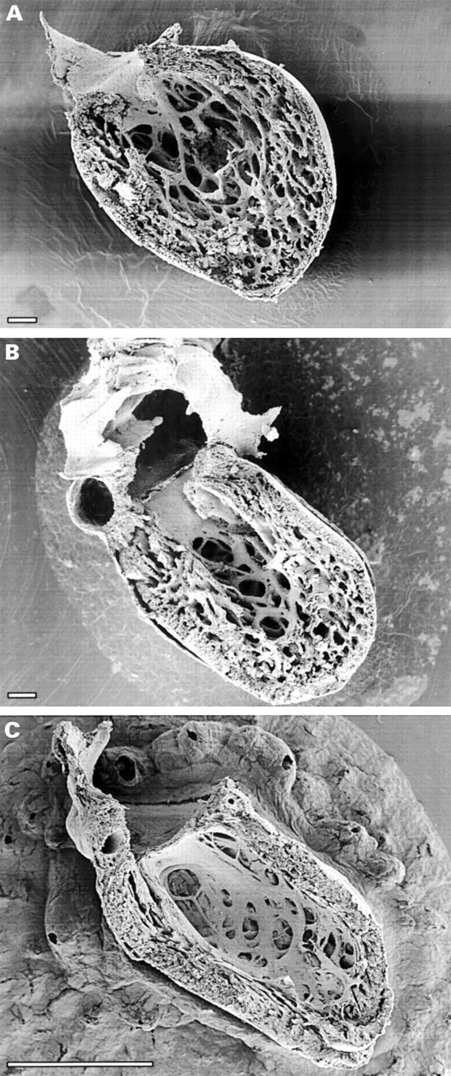
Figure 1 .

Parietal views of sagitally dissected human embryonic left ventricles showing the process of normal trabecular compaction. During early embryogenesis ventricular trabeculation develops in the apical region soon after looping, and serves primarily as a means to increase myocardial oxygenation in the absence of coronary circulation. Concomitant with ventricular septation, the trabeculae start to compact in their portions adjacent to the outer compact myocardium, adding substantially to its thickness. (A) Abundant fine trabeculae are present at six weeks. (B) The trabeculae start to solidify at their basal area, contributing to added thickness of the compact layer, at 12 weeks when ventricular septation is completed. (C) The compact layer forms most of the myocardial mass after completion of compaction in the early fetal period. Scale bars 100 µm (A, B), 1 mm (C). (A) and (C) from Sedmera et al.11


