Abstract
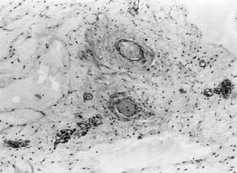
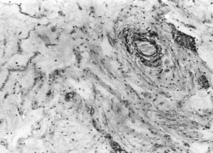
A case of aggressive angiomyxoma in a 25 year old woman is presented. The patient was admitted to hospital with a history of hesitancy of micturation and pain in the right iliac fossa. She was found to have a left labial mass, which was clinically diagnosed to be a Bartholin gland cyst. A pelvic ultrasound revealed an additional mass in the right paravesical region. At surgery, two distinct masses were removed, one from the right perivesical space and the other from the left labium. Both masses were rubbery, white, and gelatinous and showed similar histopathology findings of thick and thin walled vascular channels set in a loose myxoid stroma. A diagnosis of multifocal aggressive angiomyxoma was made. This is the first reported case of aggressive angiomyxoma occurring as two distinct masses in one patient
Key Words: aggressive angiomyxoma • myxoma • myxoid tumours • aggressive angiomyxoma of female pelvis • female perineum
Full Text
The Full Text of this article is available as a PDF (80.0 KB).
Figure 1 Section from the vulvar mass showing myxoid areas with stellate cells and blood vessels (haematoxylin and eosin stained; magnification, x100).
Figure 2 Section from the pelvic mass showing stellate cells in a myxoid stroma with prominent blood vessels (haematoxylin and eosin stained; magnification, x100).


