

Abstract
A fragment of tumour was erroneously mixed up with an endometrial biopsy. Microsatellite polymerase chain reaction (PCR) clearly demonstrated the extraneous nature of the fragment. Microsatellite PCR may be useful for the identification of mislabelled or mismatched tissue fragments in surgical pathology specimens.
Key Words: mislabelled tissue • mixed up specimens • microsatellite PCR
Full Text
The Full Text of this article is available as a PDF (118.8 KB).
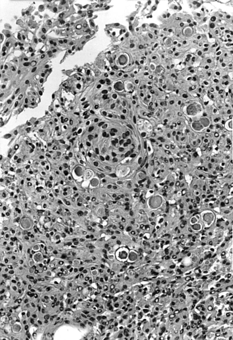
Figure 1 High power view of the presumed contaminant tissue fragment. The morphological features resemble those of a secretory meningioma.
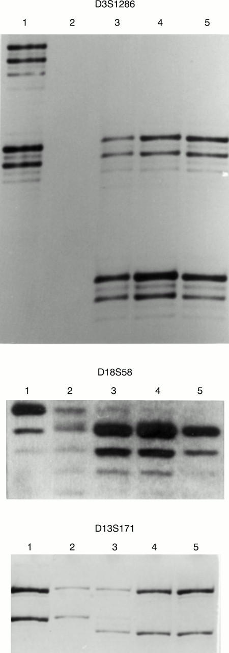
Figure 2 Representative picture of the gels corresponding to microsatellites D3S1286, D18S58, and D13SS171. Lanes 1 were DNA obtained from peripheral blood of the patient that presented with vaginal bleeding. Lanes 2 represented DNA extracted from the atrophic endometrium of the same patient as lanes 1. Lanes 3 corresponded to DNA obtained from the presumed contaminant tissue fragment. Lanes 4 represented DNA isolated from the meningioma that was interpreted as the possible source of contamination. Lanes 5 were DNA from peripheral blood of the patient with the meningioma. No amplification was obtained from DNA of the atrophic endometrium (lane 3) for marker D3S1286, because of the scarcity of the material obtained in the biopsy. The results clearly show that the suspicious fragment (lanes 3) did not match the DNA obtained from peripheral blood (lanes 1) and endometrial tissue (lanes 2) of the patient who underwent the endometrial biopsy.