Full Text
The Full Text of this article is available as a PDF (206.0 KB).
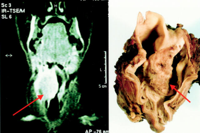
Figure 1 Coronal magnetic resonance image of a carcinoma of the right piriform fossa, seen macroscopically on the right. The specimen has been opened posteriorly to demonstrate the tumour.

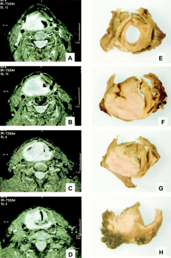
Figure 2 (A)–(D) Horizontal magnetic resonance images of the carcinoma seen in fig 1, showing invasion of the tumour along the right side of the larynx. The corresponding macroscopic slices of the larynx are shown in (E)–(H).
Selected References
These references are in PubMed. This may not be the complete list of references from this article.
- Bryne M., Koppang H. S., Lilleng R., Stene T., Bang G., Dabelsteen E. New malignancy grading is a better prognostic indicator than Broders' grading in oral squamous cell carcinomas. J Oral Pathol Med. 1989 Sep;18(8):432–437. doi: 10.1111/j.1600-0714.1989.tb01339.x. [DOI] [PubMed] [Google Scholar]
- Bryne M., Nielsen K., Koppang H. S., Dabelsteen E. Reproducibility of two malignancy grading systems with reportedly prognostic value for oral cancer patients. J Oral Pathol Med. 1991 Sep;20(8):369–372. doi: 10.1111/j.1600-0714.1991.tb00946.x. [DOI] [PubMed] [Google Scholar]
- Henson D. E. The histological grading of neoplasms. Arch Pathol Lab Med. 1988 Nov;112(11):1091–1096. [PubMed] [Google Scholar]
- Jakobsson P. A., Eneroth C. M., Killander D., Moberger G., Mårtensson B. Histologic classification and grading of malignancy in carcinoma of the larynx. Acta Radiol Ther Phys Biol. 1973 Feb;12(1):1–8. doi: 10.3109/02841867309131085. [DOI] [PubMed] [Google Scholar]
- Lumerman H., Freedman P., Kerpel S. Oral epithelial dysplasia and the development of invasive squamous cell carcinoma. Oral Surg Oral Med Oral Pathol Oral Radiol Endod. 1995 Mar;79(3):321–329. doi: 10.1016/s1079-2104(05)80226-4. [DOI] [PubMed] [Google Scholar]
- Michaels L., Gregor R. T. Examination of the larynx in the histopathology laboratory. J Clin Pathol. 1980 Aug;33(8):705–710. doi: 10.1136/jcp.33.8.705. [DOI] [PMC free article] [PubMed] [Google Scholar]
- Odell E. W., Jani P., Sherriff M., Ahluwalia S. M., Hibbert J., Levison D. A., Morgan P. R. The prognostic value of individual histologic grading parameters in small lingual squamous cell carcinomas. The importance of the pattern of invasion. Cancer. 1994 Aug 1;74(3):789–794. doi: 10.1002/1097-0142(19940801)74:3<789::aid-cncr2820740302>3.0.co;2-a. [DOI] [PubMed] [Google Scholar]
- Roland N. J., Caslin A. W., Nash J., Stell P. M. Value of grading squamous cell carcinoma of the head and neck. Head Neck. 1992 May-Jun;14(3):224–229. doi: 10.1002/hed.2880140310. [DOI] [PubMed] [Google Scholar]
- TUCKER G. F., Jr A histological method for the study of the spread of carcinoma within the larynx. Ann Otol Rhinol Laryngol. 1961 Sep;70:910–921. doi: 10.1177/000348946107000321. [DOI] [PubMed] [Google Scholar]


