

Abstract
Aims—To review the results of 73 consecutive fine needle aspirations (FNAs) that were collected by a pathologist and analysed by immunoflow cytometry. Material for a cell block was also collected from some of these lesions.
Methods—The setting was a large general hospital in rural New Zealand. The FNAs were performed by a pathologist, or a radiologist for image guided localisations. Material for immunoflow cytometry was collected into RPMI and, when required, material for a cell block was collected into formalin.
Results—Of the 73 samples collected by FNA nine were inadequate. Light chain restriction could be demonstrated in most FNA samples from B cell lymphomas (28 of 30 adequate samples). The exceptions were two cases of T cell rich B cell lymphoma. Artefactual light chain restriction was seen occasionally in T cell lymphomas, presumably as a result of autoantibodies binding to the cell surfaces. It was possible to subtype most (18 of 30 adequate samples) B cell lymphomas as chronic lymphocytic leukaemia (CLL), follicle centre cell lymphoma (FCCL), or mantle cell lymphoma. The CD4 to CD8 ratio was not usually restricted in T cell lymphomas and coexpression of CD4 and CD8 was not usually found. Loss of pan-T cell antigens was seen in some T cell lymphomas. Four of the six T cell lymphomas and three of the four non-lymphoid malignancies were diagnosed with the aid of cell block immunohistochemistry. Only one of the four cases of Hodgkin's lymphoma showed Reed-Sternberg cells in the FNA smears.
Conclusions—It is not always possible to characterise lymphomas as fully with FNA and immunoflow cytometry as is possible with biopsy histology and a full battery of modern investigations. Nevertheless, in the setting of a large rural general hospital immunoflow cytometry on FNA samples is a highly effective method of diagnosing and typing B cell lymphomas. Immunoflow cytometry is of little use for T cell lymphomas or Hodgkin's lymphomas. We advocate the use of cell block immunohistochemistry in preference to immunoflow cytometry for cases in which the cytological appearance of the specimen is overtly malignant but the differential diagnosis includes non-lymphoid malignancy.
Key Words: lymphoma • flow cytometry • cell blocks • immunolabelling • fine needle aspiration
Full Text
The Full Text of this article is available as a PDF (149.8 KB).
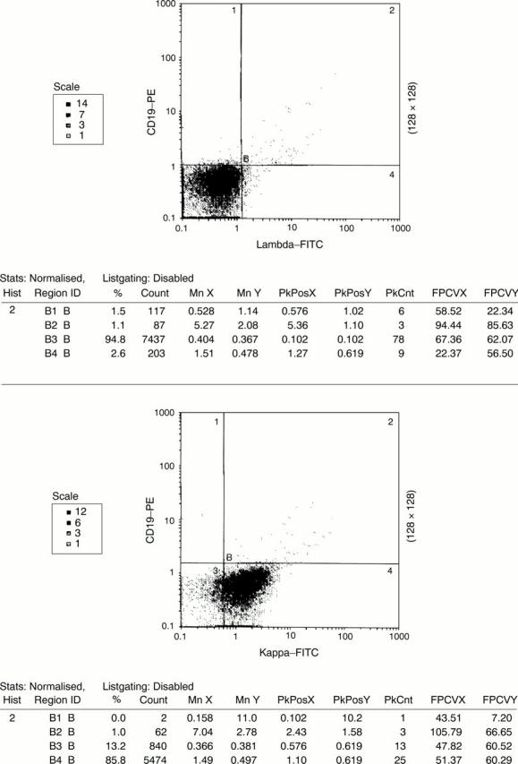
Figure 1 A scatter plot showing κ light chain restriction in a T cell lymphoma from a patient with AIDS (case 37). There was no coexpression of CD19 and κ, exemplifying the importance of dual staining in assessing light chain restriction.
Selected References
These references are in PubMed. This may not be the complete list of references from this article.
- Jeffers M. D., Milton J., Herriot R., McKean M. Fine needle aspiration cytology in the investigation on non-Hodgkin's lymphoma. J Clin Pathol. 1998 Mar;51(3):189–196. doi: 10.1136/jcp.51.3.189. [DOI] [PMC free article] [PubMed] [Google Scholar]
- Mayall F., Chang B., Darlington A. A review of 50 consecutive cytology cell block preparations in a large general hospital. J Clin Pathol. 1997 Dec;50(12):985–990. doi: 10.1136/jcp.50.12.985. [DOI] [PMC free article] [PubMed] [Google Scholar]
- Robins D. B., Katz R. L., Swan F., Jr, Atkinson E. N., Ordonez N. G., Huh Y. O. Immunotyping of lymphoma by fine-needle aspiration. A comparative study of cytospin preparations and flow cytometry. Am J Clin Pathol. 1994 May;101(5):569–576. doi: 10.1093/ajcp/101.5.569. [DOI] [PubMed] [Google Scholar]
- Sneige N., Dekmezian R. H., Katz R. L., Fanning T. V., Lukeman J. L., Ordoñez N. F., Cabanillas F. F. Morphologic and immunocytochemical evaluation of 220 fine needle aspirates of malignant lymphoma and lymphoid hyperplasia. Acta Cytol. 1990 May-Jun;34(3):311–322. [PubMed] [Google Scholar]
- Young N. A., Al-Saleem T. I., Al-Saleem Z., Ehya H., Smith M. R. The value of transformed lymphocyte count in subclassification of non-Hodgkin's lymphoma by fine-needle aspiration. Am J Clin Pathol. 1997 Aug;108(2):143–151. doi: 10.1093/ajcp/108.2.143. [DOI] [PubMed] [Google Scholar]
- Young N. A., Al-Saleem T. I., Ehya H., Smith M. R. Utilization of fine-needle aspiration cytology and flow cytometry in the diagnosis and subclassification of primary and recurrent lymphoma. Cancer. 1998 Aug 25;84(4):252–261. [PubMed] [Google Scholar]
- Zander D. S., Iturraspe J. A., Everett E. T., Massey J. K., Braylan R. C. Flow cytometry. In vitro assessment of its potential application for diagnosis and classification of lymphoid processes in cytologic preparations from fine-needle aspirates. Am J Clin Pathol. 1994 May;101(5):577–586. doi: 10.1093/ajcp/101.5.577. [DOI] [PubMed] [Google Scholar]
- Zardawi I. M., Jain S., Bennett G. Flow-cytometric algorithm on fine-needle aspirates for the clinical workup of patients with lymphadenopathy. Diagn Cytopathol. 1998 Oct;19(4):274–278. doi: 10.1002/(sici)1097-0339(199810)19:4<274::aid-dc9>3.0.co;2-a. [DOI] [PubMed] [Google Scholar]