

Abstract
Breast lymphoma accounts for less than 1% of all non-Hodgkin's lymphomas (NHLs) and approximately 0.1% of all breast neoplasms. Most breast lymphomas are classified as diffuse large B cell or mucosa associated lymphoid tissue (MALT) lymphomas. The case of a 53 year old woman presenting with a breast mass and found to have mantle cell lymphoma is described. Core biopsy of the breast lesion showed a B cell NHL, probably of large cell type and of high grade. Morphological and immunophenotypic analysis of peripheral blood and bone marrow samples suggested a mantle cell lymphoma (MCL). This was confirmed by the detection of a t(11;14) in the bone marrow aspirate and breast tissue by polymerase chain reaction analysis. There have been no previous reports of an MCL presenting as a breast lump. Because a diagnosis of MCL has prognostic and therapeutic implications, this case highlights the need for an awareness of MCL presenting in this way, and the requirement for specialised investigations in its detection.
Key Words: mantle cell lymphoma • breast lymphoma • cyclin D1 • t(11;14)
Full Text
The Full Text of this article is available as a PDF (203.5 KB).
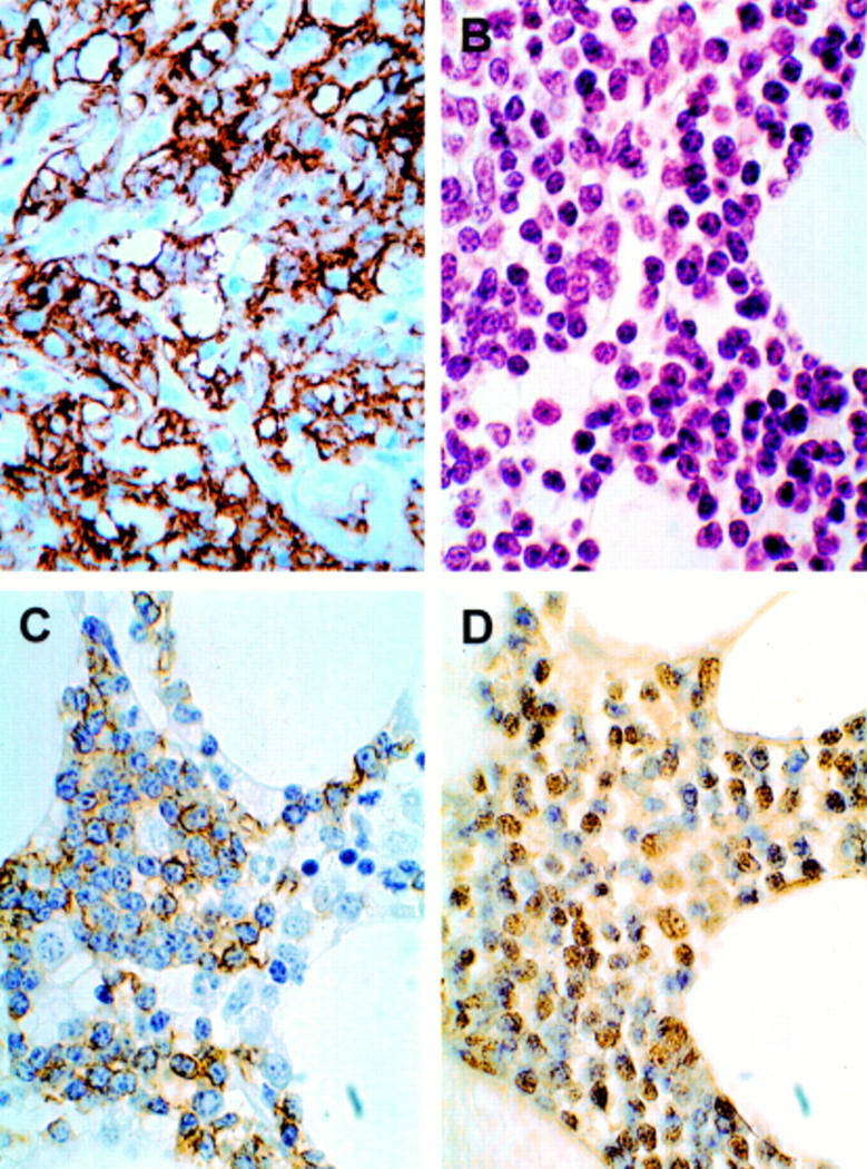
Figure 1 (A) Breast core biopsy. Lymphoma cells showing membrane staining for CD20. Immunoperoxidase, haematoxylin counterstain; original magnification, x400. (B) Bone marrow trephine. Infiltrate of intermediate sized centrocyte-like malignant lymphoid cells. Haematoxylin and eosin; original magnification, x630. (C) Bone marrow trephine. Membrane staining of neoplastic lymphoid cells for CD5. Immunoperoxidase, haematoxylin counterstain; original magnification, x630. (D) Bone marrow trephine. Nuclear staining in neoplastic lymphoid cells for cyclin D1. Immunoperoxidase, haematoxylin counterstain; original magnification, x630.

Figure 2 PCR amplified products from bcl-1/JH consensus primers visualised with ethidium bromide in a UV illuminated agarose gel. Lane 1, negative control without DNA; lane 2, positive t(11;14) control; lane 3, negative control with DNA; lane 4, amplified DNA from patient; lane M, molecular weight markers.