Abstract
Aims—Prolactin plays an important role in the proliferation and differentiation of normal breast epithelium, and possibly in the development of breast carcinoma. The effects of prolactin are mediated by its receptor; thus, alteration in the expression of this receptor could be important in studying the biology of breast cancer. This investigation was aimed at comparing the expression of prolactin receptors in normal, benign, and malignant breast tissue.
Material/Methods—The expression of prolactin receptors was studied in paraffin wax embedded sections of 102 breast biopsies (93 female and nine male), using the monoclonal antibody B6.2, and the avidin–biotin immunoperoxidase technique. Six biopsies were normal, 34 had benign lesions, and 62 were malignant.
Results—In normal cases, prolactin receptor positivity was seen only on the luminal borders of the epithelial cells lining ducts and acini. In most benign lesions, variable degrees of luminal and cytoplasmic staining were seen. Cells showing apocrine metaplasia and florid regular ductal epithelial hyperplasia were mostly negative. In malignant cases, the staining pattern was mostly cytoplasmic and heterogeneous. Forty one of the 59 carcinomas in women showed a degree of positivity involving 10–100% of the tumour cells. A significant direct correlation was found between prolactin receptor and oestrogen receptor staining when only cases that scored more than 100/300 for the latter receptor, using the H scoring system, were considered (p = 0.0207). No correlation was found between prolactin receptors and progesterone receptors, patient's age, tumour size, tumour grade, or axillary lymph node status.
Conclusions—Prolactin receptors seem to be expressed at different cellular sites in normal, benign, and malignant breast epithelial cells. The receptor is expressed in more than two thirds of female breast carcinomas, suggesting that it may play a role in the pathogenesis of the disease. The positivity is correlated with moderate and strong staining for oestrogen receptors in tissue sections, but not with other prognostic factors.
Key Words: breast • breast carcinoma • male breast • prolactin receptors • oestrogen receptors
Full Text
The Full Text of this article is available as a PDF (194.8 KB).
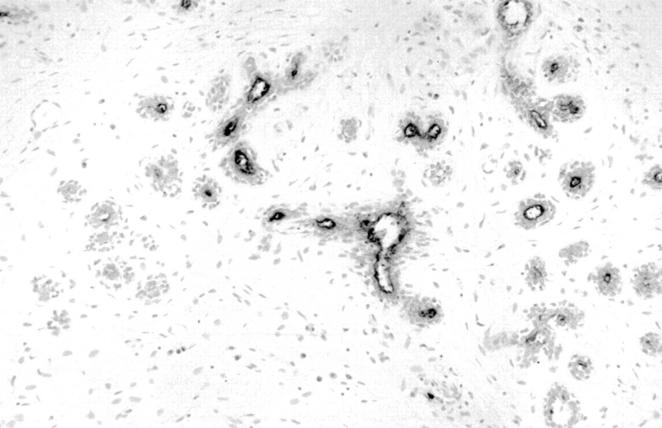
Figure 1 Normal mammary ducts and acini showing prolactin receptor luminal staining.
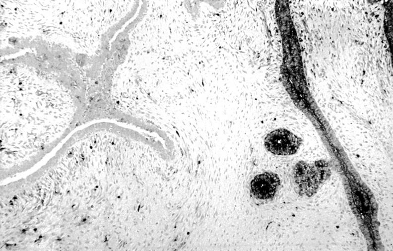
Figure 2 Fibroadenoma, prolactin receptor positive zonal cytoplasmic staining. Scattered positively stained lymphocytes are also seen.
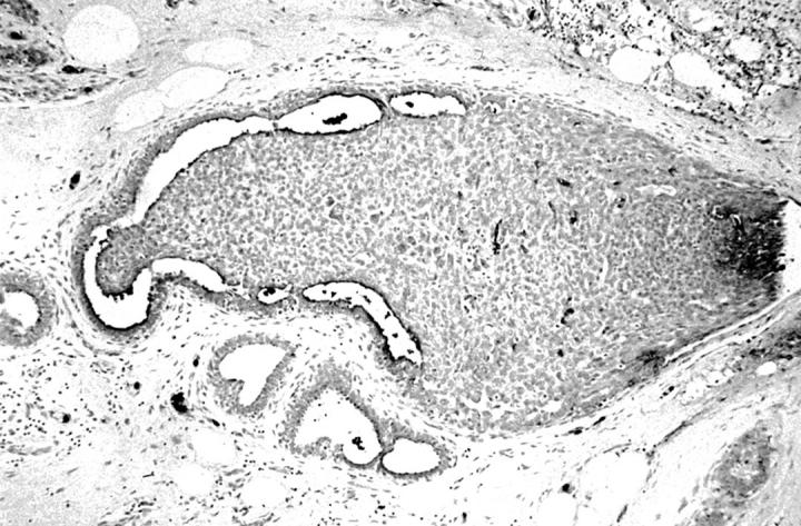
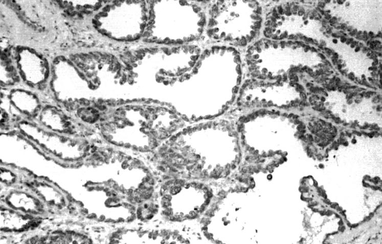
Figure 3 Florid ductal epithelial hyperplasia, mostly prolactin receptor negative, except for focal luminal staining.
Figure 4 Lactational adenoma, positive prolactin receptor cytoplasmic staining.
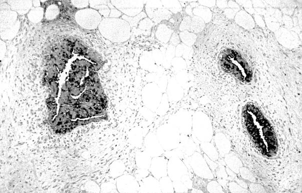
Figure 5 Gynaecomastia, strong positive prolactin receptor luminal and cytoplasmic staining.
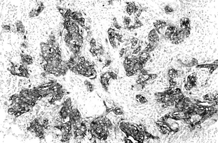
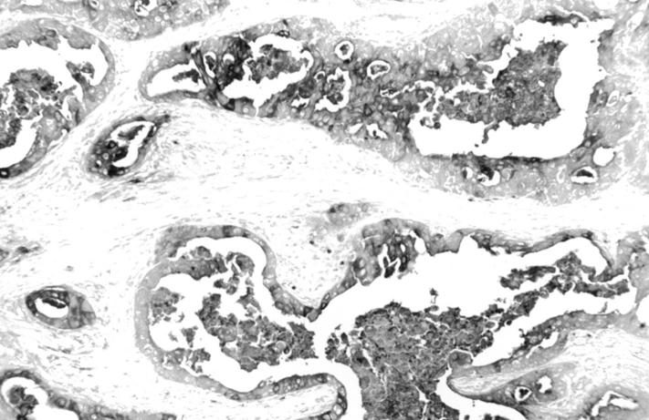
Figure 6 Invasive ductal carcinoma, showing strongly positive cytoplasmic and membrane staining for the prolactin receptor. Note unstained stroma between tumour cells.
Figure 7 Invasive apocrine carcinoma showing focal positive cytoplasmic and luminal staining for the prolactin receptor.
Selected References
These references are in PubMed. This may not be the complete list of references from this article.
- Banerjee R., Ginsburg E., Vonderhaar B. K. Characterization of a monoclonal antibody against human prolactin receptors. Int J Cancer. 1993 Nov 11;55(5):712–721. doi: 10.1002/ijc.2910550503. [DOI] [PubMed] [Google Scholar]
- Ben-Jonathan N., Mershon J. L., Allen D. L., Steinmetz R. W. Extrapituitary prolactin: distribution, regulation, functions, and clinical aspects. Endocr Rev. 1996 Dec;17(6):639–669. doi: 10.1210/edrv-17-6-639. [DOI] [PubMed] [Google Scholar]
- Bole-Feysot C., Goffin V., Edery M., Binart N., Kelly P. A. Prolactin (PRL) and its receptor: actions, signal transduction pathways and phenotypes observed in PRL receptor knockout mice. Endocr Rev. 1998 Jun;19(3):225–268. doi: 10.1210/edrv.19.3.0334. [DOI] [PubMed] [Google Scholar]
- Bonneterre J., Peyrat J. P., Beuscart R., Lefebvre J., Demaille A. Prognostic significance of prolactin receptors in human breast cancer. Cancer Res. 1987 Sep 1;47(17):4724–4728. [PubMed] [Google Scholar]
- Boutin J. M., Jolicoeur C., Okamura H., Gagnon J., Edery M., Shirota M., Banville D., Dusanter-Fourt I., Djiane J., Kelly P. A. Cloning and expression of the rat prolactin receptor, a member of the growth hormone/prolactin receptor gene family. Cell. 1988 Apr 8;53(1):69–77. doi: 10.1016/0092-8674(88)90488-6. [DOI] [PubMed] [Google Scholar]
- Clevenger C. V., Chang W. P., Ngo W., Pasha T. L., Montone K. T., Tomaszewski J. E. Expression of prolactin and prolactin receptor in human breast carcinoma. Evidence for an autocrine/paracrine loop. Am J Pathol. 1995 Mar;146(3):695–705. [PMC free article] [PubMed] [Google Scholar]
- De Placido S., Gallo C., Perrone F., Marinelli A., Pagliarulo C., Carlomagno C., Petrella G., D'Istria M., Delrio G., Bianco A. R. Prolactin receptor does not correlate with oestrogen and progesterone receptors in primary breast cancer and lacks prognostic significance. Ten year results of the Naples adjuvant (GUN) study. Br J Cancer. 1990 Oct;62(4):643–646. doi: 10.1038/bjc.1990.346. [DOI] [PMC free article] [PubMed] [Google Scholar]
- FOULDS L. The experimental study of tumor progression: a review. Cancer Res. 1954 Jun;14(5):327–339. [PubMed] [Google Scholar]
- García-Caballero T., Morel G., Gallego R., Fraga M., Pintos E., Gago D., Vonderhaar B. K., Beiras A. Cellular distribution of prolactin receptors in human digestive tissues. J Clin Endocrinol Metab. 1996 May;81(5):1861–1866. doi: 10.1210/jcem.81.5.8626848. [DOI] [PubMed] [Google Scholar]
- Hanna W., Kahn H. J. Ultrastructural and immunohistochemical characteristics of mucoepidermoid carcinoma of the breast. Hum Pathol. 1985 Sep;16(9):941–946. doi: 10.1016/s0046-8177(85)80133-7. [DOI] [PubMed] [Google Scholar]
- Hennighausen L., Robinson G. W., Wagner K. U., Liu W. Prolactin signaling in mammary gland development. J Biol Chem. 1997 Mar 21;272(12):7567–7569. doi: 10.1074/jbc.272.12.7567. [DOI] [PubMed] [Google Scholar]
- Kline J. B., Roehrs H., Clevenger C. V. Functional characterization of the intermediate isoform of the human prolactin receptor. J Biol Chem. 1999 Dec 10;274(50):35461–35468. doi: 10.1074/jbc.274.50.35461. [DOI] [PubMed] [Google Scholar]
- Matera L., Geuna M., Pastore C., Buttiglieri S., Gaidano G., Savarino A., Marengo S., Vonderhaar B. K. Expression of prolactin and prolactin receptors by non-Hodgkin's lymphoma cells. Int J Cancer. 2000 Jan 1;85(1):124–130. doi: 10.1002/(sici)1097-0215(20000101)85:1<124::aid-ijc22>3.0.co;2-u. [DOI] [PubMed] [Google Scholar]
- Mertani H. C., Garcia-Caballero T., Lambert A., Gérard F., Palayer C., Boutin J. M., Vonderhaar B. K., Waters M. J., Lobie P. E., Morel G. Cellular expression of growth hormone and prolactin receptors in human breast disorders. Int J Cancer. 1998 Apr 17;79(2):202–211. doi: 10.1002/(sici)1097-0215(19980417)79:2<202::aid-ijc17>3.0.co;2-b. [DOI] [PubMed] [Google Scholar]
- Nandi S., Guzman R. C., Yang J. Hormones and mammary carcinogenesis in mice, rats, and humans: a unifying hypothesis. Proc Natl Acad Sci U S A. 1995 Apr 25;92(9):3650–3657. doi: 10.1073/pnas.92.9.3650. [DOI] [PMC free article] [PubMed] [Google Scholar]
- Ormandy C. J., Hall R. E., Manning D. L., Robertson J. F., Blamey R. W., Kelly P. A., Nicholson R. I., Sutherland R. L. Coexpression and cross-regulation of the prolactin receptor and sex steroid hormone receptors in breast cancer. J Clin Endocrinol Metab. 1997 Nov;82(11):3692–3699. doi: 10.1210/jcem.82.11.4361. [DOI] [PubMed] [Google Scholar]
- Partridge R. K., Hähnel R. Prolactin receptors in human breast carcinoma. Cancer. 1979 Feb;43(2):643–646. doi: 10.1002/1097-0142(197902)43:2<643::aid-cncr2820430235>3.0.co;2-c. [DOI] [PubMed] [Google Scholar]
- Peters F., Schuth W. Hyperprolactinemia and nonpuerperal mastitis (duct ectasia). JAMA. 1989 Mar 17;261(11):1618–1620. [PubMed] [Google Scholar]
- Peters F., Schuth W., Scheurich B., Breckwoldt M. Serum prolactin levels in patients with fibrocystic breast disease. Obstet Gynecol. 1984 Sep;64(3):381–385. [PubMed] [Google Scholar]
- Reynolds C., Montone K. T., Powell C. M., Tomaszewski J. E., Clevenger C. V. Expression of prolactin and its receptor in human breast carcinoma. Endocrinology. 1997 Dec;138(12):5555–5560. doi: 10.1210/endo.138.12.5605. [DOI] [PubMed] [Google Scholar]
- Rowe P. H. Granulomatous mastitis associated with a pituitary prolactinoma. Br J Clin Pract. 1984 Jan;38(1):32–34. [PubMed] [Google Scholar]
- Selim A. G., Wells C. A. Immunohistochemical localisation of androgen receptor in apocrine metaplasia and apocrine adenosis of the breast: relation to oestrogen and progesterone receptors. J Clin Pathol. 1999 Nov;52(11):838–841. doi: 10.1136/jcp.52.11.838. [DOI] [PMC free article] [PubMed] [Google Scholar]
- Shousha S., Backhouse C. M., Dawson P. M., Alaghband-Zadeh J., Burn I. Mammary duct ectasia and pituitary adenomas. Am J Surg Pathol. 1988 Feb;12(2):130–133. doi: 10.1097/00000478-198802000-00006. [DOI] [PubMed] [Google Scholar]
- Shousha S. Breast carcinoma presenting during or shortly after pregnancy and lactation. Arch Pathol Lab Med. 2000 Jul;124(7):1053–1060. doi: 10.5858/2000-124-1053-BCPDOS. [DOI] [PubMed] [Google Scholar]


