Abstract
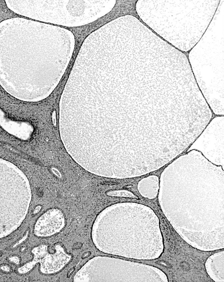
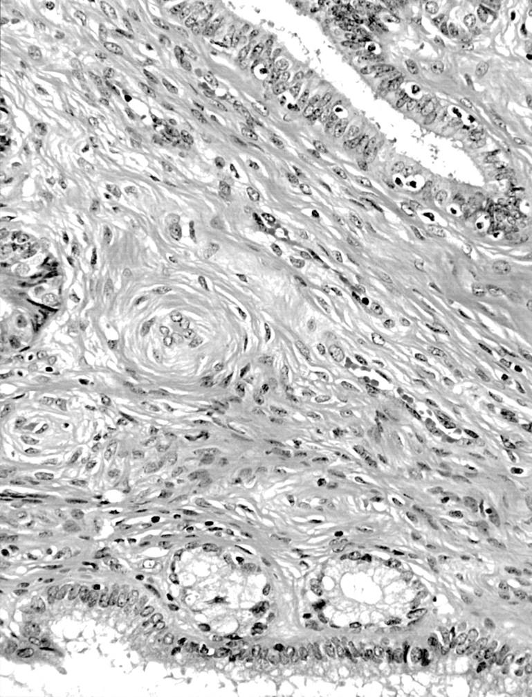
A 73 year old woman presented with a right sided adnexal cystic mass. At laparotomy, this proved to be a benign serous ovarian cyst and an aggregation of thin walled subserosal and soft tissue cysts and spongy nodules up to 16 mm in diameter involving the side wall of the uterus and adjacent parametrium. These were removed by total abdominal hysterectomy and bilateral salpingo-oophorectomy. Histologically, the cystic spaces and smaller acini were lined by benign tubo-endometrioid epithelium, with smaller areas typical of serous differentiation and rare microfoci of endocervical-type mucinous epithelium. These features indicated multidirectional Mullerian differentiation in a process that, overall, was consistent with so called florid cystic endosalpingiosis. This lesion is to be distinguished from other benign conditions including multicystic mesothelioma, endometriosis, endocervicosis, florid deep glands of the uterine cervix, and deep Nabothian cysts of the uterine cervix.
Key Words: uterus • endosalpingiosis • cysts
Full Text
The Full Text of this article is available as a PDF (106.7 KB).

Figure 1 Florid cystic endosalpingiosis. Variably sized cystic spaces are lined by tubo-endometrial and flattened more serous epithelium. Haematoxylin and eosin stained; original magnification, x40.

Figure 2 Florid cystic endosalpingiosis. Variable differentiation is demonstrated with spaces lined partly by mucinous and tubal type epithelium. Original magnification, x40.


