

Abstract
A 60 year old woman who presented with dysphagia and weight loss was found to have multiple foci of dysplasia and in situ and invasive squamous cell carcinoma scattered along the whole length of the oesophagus, with intervening areas of normal mucosa. The patient had a history of two breast carcinomas 19 and one year previously for which she had repeated radiotherapy. Several members of the patient's close family had histories of malignant disease. All oesophageal lesions and the more recent breast cancer showed positive immunostaining for p53 protein. p53 mutations, some involving different exons, were also detected in these lesions. No p53 immunostaining or mutations were detected in the normal oesophageal mucosa. The findings suggest an independent origin of the multiple dysplastic and neoplastic foci, which might have developed in a background of a field change, possibly related to the previous radiotherapy. The strong family history of malignant diseases raises the possibility that, in addition, genetic factors might have played a role in the development of the oesophageal disease.
Key Words: oesophageal carcinoma • breast carcinoma • p53 • radiotherapy
Full Text
The Full Text of this article is available as a PDF (135.0 KB).
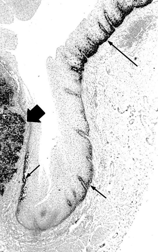
Figure 1 Positive p53 immunostaining of a focus of primary invasive squamous cell carcinoma (thick arrow), as well as scattered small foci of dysplasia (thin arrows). Note intervening p53 negative, non-stained, normal mucosa. Immunoperoxidase stain; original magnification, x16.