Abstract
We report the engineering of the surface of the tobacco mosaic virus (TMV) virion with a mosquito decapeptide hormone, trypsin-modulating oostatic factor (TMOF). The TMV coat protein (CP) was fused to TMOF at the C terminus by using a read-through, leaky stop codon that facilitated expression of CP and chimeric CP-TMOF (20:1 ratio) that were coassembled into virus particles in infected Nicotiana tabacum. Plants that were infected with the hybrid TMV RNA accumulated TMOF to levels of 1.3% of total soluble protein. Infected tobacco leaf discs that were fed to Heliothis virescens fourth-instar larvae stunted their growth and inhibited trypsin and chymotrypsin activity in their midgut. Purified CP-TMOF virions fed to mosquito larvae stopped larval growth and caused death. Because TMV has a wide host range, expressing TMV-TMOF in plants can be used as a general method to protect them against agricultural insect pests and to control vector mosquitoes.
Keywords: genetic engineering, Helitothis virescens, mosquitoes, plants, digestion
The major digestive enzymes of many insect species are midgut serine proteases, including trypsin and chymotrypsin. Thus, controlling digestion in insects by using trypsin inhibitors has received substantial attention because of their widespread occurrence and potential for insect control (1–4). In anautogenous adult mosquitoes and their larvae, trypsin and chymotrypsin are the major gut digestive enzymes. These enzymes digest either the protein-rich blood meal the adult female uses for egg development (5) or proteinacious food from the water for larval growth and development (6). The sequence of physiological events that follows the uptake of a blood meal in female mosquitoes or protein uptake in larvae is complicated, involving osmotic pressure, juvenile hormone III, and other unknown factors (5). Similar to mosquito larvae, protein digestion in the tobacco budworm (Heliothis virescens) is mediated by endo- and exopeptidases secreted from the midgut epithelium cells into the luminal fluid of the midgut (7). The main digestive enzymes in H. virescens midgut are the serine proteases (8). Because mosquitoes transmit many diseases, such as malaria, dengue, and yellow fever, that exert social and economical burden in tropical countries, and H. virescens causes extensive agricultural damage, there is a considerable interest to control these insects.
Although the factor(s) that directly stimulates trypsin biosynthesis in mosquito and H. virescens are not known, a decapeptide (YDPAPPPPPP) named trypsin-modulating oostatic factor (TMOF) that terminates trypsin biosynthesis in mosquitoes and H. virescens has been identified and characterized (9, 10). Mosquito ovaries synthesize and release TMOF into the hemolymph after a blood meal (11), and the hormone modulates the synthesis of trypsin in the mosquito gut by binding a TMOF receptor (12). Cytoimmunochemical studies showed that TMOF is also found in the brain and the neuroendocrine organs of adult and larval Aedes aegypti (13). Thus, the hormone has a dual role in terminating serine-proteases biosynthesis not only in adult female mosquitoes but also in larvae (13). Mosquito TMOF or its analogues stop trypsin biosynthesis in the cat flea Ctenocephalides felis, in the stable fly Stomoxys calcitrans, in the house fly Musca domestica, in the midge Culicoides variipenis, and in H. virescens (8–10). TMOF from the gray fleshfly Neobellieria bullata (Neb-TMOF) has been sequenced and characterized. The hormone is an unblocked hexapeptide (NPTNLH), that, like A. aegypti TMOF (Aea-TMOF), stops trypsin biosynthesis and egg development in the fleshfly (14). In the fleshfly, it was shown that Neb-TMOF controls the translation but not the transcription of the trypsin gene (15).
Because TMOF affects the synthesis of trypsin-like enzymes in the larval gut of mosquito and H. virescens, we decided to explore the possibility of fusing TMOF with the coat protein (CP) of tobacco mosaic virus (TMV), which produces large quantities of RNA and protein in infected plant cells. TMV causes very small degree of mosaic symptoms and only slight reduction in plant growth after the genetic modification to the CP (16). Although TMV is known to exist on human skin, it does not infect humans. A hybrid viral RNA containing the TMOF sequence fused to the coat protein ORF of TMVU1 (17) was constructed so that 5% of the CP subunits had TMOF fused to the carboxyl terminus. Virus particles isolated from plants infected with this chimeric virus presented TMOF on their surface. The capacity to engineer crops to synthesize insect peptide hormones could be used in the future to protect crops against agricultural pest insects and to harvest peptides that can be used to control mosquito larvae.
Results
Bioengineering of Chimeric TMV-Aea-TMOF.
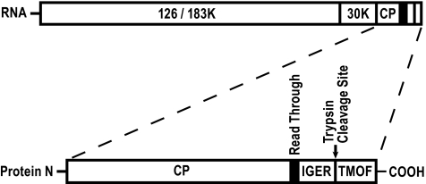
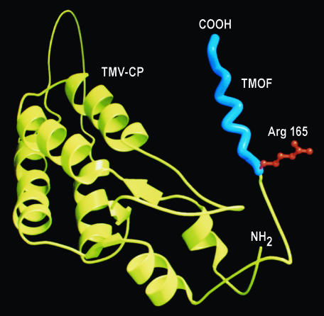
A hybrid TMV RNA was constructed containing a replicase (126/183 kDa protein) and a cell to cell movement protein (30 kDa) ORF and the CP ORF with a read-through sequence (5′-TAGCAATTA-3′) followed by a trypsin cleavage site (IGER) fused to the Aea-TMOF. The leaky stop signal allowed a virion assembly with a predicted ratio of 1:20 (CP-TMOF:CP) (Fig. 1) (18). A bioengineered TMV without the leaky stop signal transcribed TMOF on every viral CP but did not assemble into virus particles because of steric hindrance between every CP caused by the left-handed helical conformation of TMOF (Fig. 2) (18).
Fig. 1.
Genome organization of TMV-TMOF containing a read-through sequence. TMV-U1 ORF representing the replicase of 126 and 183 kDa (126K/183K), the cell-to-cell movement protein 30 kDa (30K), and the viral coat protein (CP) was fused to a read-through sequence (5′-TAGCAATTA-3′), a trypsin cleavage site (IEGR), and an A. aegypti TMOF (YDPAPPPPPP) sequence behind a T7 promoter at the 5′ end of the construct. The TMV-TMOF RNA was transcribed by using T7 RNA polymerase, and the transcribed RNA was mechanically inoculated onto Ni. tabacum (L) cv. xanthu. Gene distances are not proportional to gene sizes.
Fig. 2.
Ribbon drawing by means of MOLSCRIPT of TMV coat protein (CP) fused to A. aegypti TMOF. The TMV-CP is shown in yellow, and the N terminus is shown as NH2. TMOF exhibiting a left-handed helix is shown in blue with a trypsin cleavage site at Arg165 in red. The C terminus of the fusion protein is at the carboxyl end of TMOF (COOH).
Characterization of CP-TMOF.
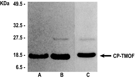
Chimeric TMV-TMOF particles were purified from infected tobacco leaves 2 weeks after inoculation. Protein samples from 22 and 28 μg of isolated virions, respectively, were fractionated by 12% SDS/PAGE and subjected to Western blot analysis with antibody to the CP protein. CP antibody recognized CP-TMOF protein band at 18.5 kDa (Fig. 3, lanes A and B) that comigrated with purified wild-type viral CP (17.5 kDa; Fig. 3, lane C) and a faint band at 27.5 kDa, which is an aggregation of the CP and the CP-TMOF that was not completely denatured (Fig. 3, lanes B and C). Because the mobility of the wild-type CP (17.5 kDa) and the chimeric CP-TMOF (18.5 kDa) band on 12% SDS/PAGE is the same, the CP-TMOF was further analyzed. Treatment of the chimeric TMV-TMOF with trypsin-liberated TMOF that then was analyzed by HPLC interfaced to electrospray ionization (ESI) on a triple quadrapole mass spectrometer. Abundant (M + H)+ and (M + 2H)2+ ions were observed at m/z 1,047 and 524, respectively. A collision-activated dissociation spectrum recorded on the (M + 2H)2+ ions confirmed the sequence YDPAPPPPPP, as was described in refs. 9 and 10. When synthetic TMOF (24 pmol) was added to the trypsin digest, and the resulting sample was analyzed by HPLC ESI/MS, signals observed previously at m/z 1,047 and 524 increased by a factor of 4. We conclude that the TMOF expressed in the chimeric TMV-TMOF and released with trypsin has an identical mass spectra to synthetic TMOF and has the sequence YDPAPPPPPP, as was described in refs. 9 and 10. Wild-type CP (control) did not liberate TMOF after incubation with trypsin and mass spectra analysis. The purified chimeric viral particles (396 μg) analyzed by ELISA showed that the amount of TMOF presented on the surface of the chimeric TMV was 1.189 ± 0.11 (micrograms ± SEM; n = 3), which is in good agreement with the predicted value of 1.188 μg calculated for a ratio 1:20 (CP-TMOF:CP).
Fig. 3.
Western blot analysis and SDS/PAGE of CP-TMOF. Purified TMV-TMOF virions (22 μg, lane A; 28 μg, lane B) were run on SDS/PAGE, blotted to a membrane, and analyzed by antiserum against TMV-CP. (Lane C) Wild-type TMV-CP virions (28 μg) were run on SDS/PAGE and stained with Coomassie brilliant blue for comparison. Arrow indicates the comigration of the CP (17.5 kDa) and CP-TMOF (18.5 kDa).
Effect of Feeding CP-TMOF to Mosquito Larvae.
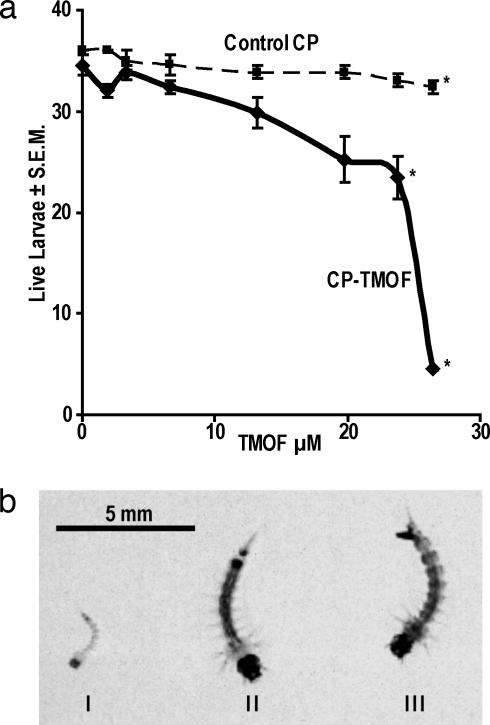
Mosquito larvae are aquatic animals in which the concentration of solute in the hemolymph is higher than in water. As a consequence, they do not drink, and factors that are dissolved in water will not enter the gut unless the larvae ingest solid food particles and at the same time swallow some water. Because mosquito larvae are filter feeders (6), we adsorbed the CP-TMOF virions on dried yeast to allow the larvae to ingest it. Feeding A. aegypti larvae (36 per group) CP-TMOF virions (0–2.33 μg/μl) containing TMOF (0–26.4 μM) caused a significant increase (n = 3; P < 0.05) in larval mortality from 0% to 4% in controls that were fed yeast or CP virions, to 35% and 87.5% in larvae that were fed CP-TMOF virions (23.7 and 26.4 μM TMOF, respectively) (Fig. 4a). Larvae that were fed on CP-TMOF virions (26.4 μM TMOF) did not grow and starved to death, whereas larvae that were fed CP or yeast grew normally (Fig. 4b). Larvae that were fed CP-TMOF virions (26.4 μM TMOF) were 2.38-fold shorter (P < 0.006; n = 20) than controls that were fed yeast (2.8 ± 0.16 and 6.66 ± 0.084 mm ± SEM, respectively), and trypsin activity in the gut of these larvae was 90% inhibited (results not shown). These results confirm an earlier report that TMOF adsorbed onto yeast particles inhibits trypsin activity in A. aegypti and Culex quinquefasciatus larvae (13). No difference in length or inhibition of trypsin activity was found when CP virions or yeast cells were fed to larvae. Thus, the inhibition of larval growth was caused by TMOF.
Fig. 4.
Feeding A. aegypti larvae CP-TMOF virions. (a) Three groups of first-instar larvae were fed increasing concentrations of CP-TMOF virions in the presence of Brewer's yeast for 6 days (solid line). Control groups were fed CP virions without TMOF (dashed line). Larval survival was followed daily and plotted against TMOF concentrations (micromolar) calculated from the concentrations of CP-TMOF virions that were fed. The results are expressed as means of three determinations ± SEM, and the experiment was repeated three times. Significant differences: ∗, P < 0.005 in larval mortality between feeding CP-TMOF virions and CP virions (control). (b) Effect on A. aegypti larval growth after feeding larvae CP-TMOF virions (I), CP virions (II), and Brewer's yeast (III) for 6 days. (Scale bar: 5 mm.)
Feeding Tobacco Leaves Infected with TMV-TMOF to H. virescens.
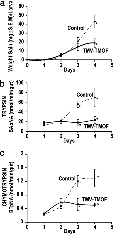
Feeding 24 fourth-instar H. virescens larvae weighing 18–19 mg tobacco leaf discs infected with TMV-TMOF for 4 days caused 2.3-fold reduction in weight (n = 6; P < 0.009) as compared with noninfected controls (Fig. 5a). ANOVA showed that there were significant differences as a result of TMOF, F(1, 37) = 8.68, P < 0.0055; larval age, F(3, 37) = 28.64, P < 0.0001; and the interaction of age, and TMOF treatment, F(3, 37) = 5.05, P < 0.005. Post hoc Tukey honestly selectively different (HSD) analyses revealed that larvae that were fed TMV-TMOF had significantly lower weight gain on day 4, and larval weight did not change significantly during the first 3 days of feeding, indicating that TMV-TMOF prevented the growth of H. virescens. Analyzing larval midguts showed that trypsin activity in larvae that were fed TMV-TMOF was reduced 3.1-fold (n = 6; P < 0.001) at day 3 and 2.2-fold (n = 6; P < 0.037) at day 4 (Fig. 5b). ANOVA showed that there were significant differences as a result of TMOF, F(1, 34) = 15.91, P < 0.0003; larval age, F(3, 34) = 8.18, P < 0.0003; and the interaction of age and TMOF treatment, F(3, 34) = 6.69, P < 0.001. Post hoc Tukey HSD analyses revealed that larvae that were fed TMV-TMOF had significantly lower trypsin activity on days 3 and 4, and trypsin activity did not change over the 4-day period. Similarly, chymotrypsin activity in the midguts was reduced 1.5-fold (n = 6; P < 0.014) at day 3 and 2.6-fold (n = 4; P < 0.046) at day 4 (Fig. 5c). ANOVA showed that there were significant differences as a result of TMOF, F(1, 23) = 10.86, P < 0.0032; larval age, F(3, 23) = 8.24, P < 0.0007; and the interaction of age and TMOF treatment, F(3, 23) = 5.32, P < 0.0062. Post hoc Tukey HSD analyses revealed that larvae that were fed TMV-TMOF had lower chymotrypsin activities on days 3 and 4, and the chymotrypsin activity of the TMV-TMOF fed larvae did not change over the 4-day period. The experiment was repeated two more times with similar results (data not shown). These results indicate that TMV-TMOF causes inhibition of trypsin and chymotrypsin activity in the midgut of H. virescens preventing normal larval growth.
Fig. 5.
Effect of feeding H. virescens tobacco leaf discs that were infected with TMV-TMOF. (a) Weight gain, after feeding for 4 days on noninfected (control). ∗, P < 0.009 versus TMV-TMOF infected leaf discs. (b) Trypsin activity in the gut after feeding for 3 and 4 days on noninfected (control). ∗, P < 0.001 and P < 0.037, respectively, versus on TMV-TMOF-infected disc leaves. (c) Chymotrypsin activity in the gut after feeding for 3 and 4 days on noninfected. ∗, P < 0.014 and P < 0.046, respectively, versus TMV-TMOF-infected leaf discs. Dashed line, larvae fed on control leaf discs; solid line, larvae fed on leaf discs infected with TMV-TMOF. Each point represents a mean ± SEM of individual larvae (n = 4–6), and the results represent one of three independent experiments.
Effect of TMOF on the Trypsin Gene.
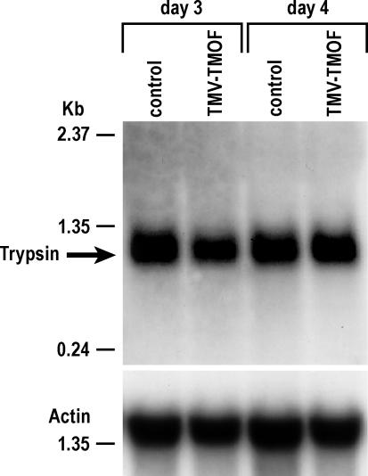
Larvae were fed TMV-TMOF infected or noninfected leaf discs for 3 and 4 days, and their guts were dissected and tested for H. virescens trypsin transcript by Northern blot analysis. Trypsin gene transcript levels were equal in the guts of larvae that were fed noninfected (control) or infected TMV-TMOF leaf discs for 3 and 4 days (Fig. 6), although trypsin activity was reduced 3.1- and 2.1-fold after 3 and 4 days of feeding the CP-TMOF virions to larvae, respectively (Fig. 5b). These results indicate that TMOF affects the translation of the trypsin message in the gut.
Fig. 6.
Northern blot analysis of H. virescens larval gut-trypsin transcript after feeding for 3 and 4 days on leaf discs that were not infected (control) and infected with TMV-TMOF. The blot was probed with trypsin and actin probes. Arrow represents expected trypsin transcript at ≈0.9 Kb. The Northern blot analysis was repeated twice.
Discussion
We constructed virions of TMVU1 (19) to present TMOF on the surface by using a read-through sequence so that the TMOF peptide was presented from the C-terminal of 5% of the viral CP subunits. This approach allowed efficient assembly of the viral CP subunits despite the steric hindrance that the left-handed helical portion of TMOF probably exerted. When it was expressed on each CP subunit virion assembly was prevented (unpublished observations). The same strategy was used successfully in producing malarial epitopes and angiotensin I-converting enzyme inhibitor on the CP of TMV. In both cases, the recombinant CP and the wild-type CP coassembled into virus particles only after a leaky stop signal was used with a ratio of 1:20 (chimeric CP:CP) (18, 20). Turpen et al. (18) reported that 62% of the coat protein is unavailable for substitutions because of steric hindrance. Thus, these reports combined with our observations suggest that expressing recombinant proteins on each CP prevents assembly of the viral particles by steric hindrance. The release of TMOF in the larval gut from the virions by trypsin allowed a rapid transport of the free TMOF through the gut epithelial cells into the hemolymph (21). In the hemolymph, the hormone bound its gut receptor located at the hemolymph side of the gut (12) and caused cessation of trypsin activity in the gut, starvation, and eventual death (13). This mode of action is different from Bacillus thuringiensis toxins that bind to receptors located in the larval midgut epithelial brush border membrane and insert into the membrane, causing formation of pores or ion channels and osmotic pressure imbalance that eventually kills the insects (22, 23).
The ability of the CP-TMOF chimera to starve and kill mosquito larvae was demonstrated when CP-TMOF virions were adsorbed onto yeast particles and fed to A. aegypti larvae for 5–6 days. The estimated lethal dose of TMOF that fused to the CP and caused 87.5% mortality was ≈26.4 μM (equivalent to 140 ng/μl TMOF or 2.33 μg/μl CP-TMOF), this concentration is 7.5-fold lower than when TMOF alone is adsorbed onto yeast particles and fed to larvae (13). It is possible that the CP-TMOF virions bind better to the yeast particles or that the CP-TMOF virions aggregate and then bind to the yeast cells, making it easier for the larvae to internalize (6). Lower concentrations of TMOF caused less mortality because at lower concentrations, less CP-TMOF virions bound to the yeast particles that the mosquito larvae fed on; mosquito larvae are filter feeders and swallow particles (6). When radioactively labeled TMOF was incubated with the same amount of yeast particles that were fed to larval mosquito, ≈10% of the radioactively labeled TMOF bound the yeast particles (D.B., unpublished observations). Using these results, we estimate that ≈10% of the available CP-TMOF virions also would bind the yeast particles that were added to each well; this amount of yeast is sufficient for larval development (13). Thus, the effective concentrations that caused high mortality are probably 10-fold lower than the total concentrations that were used during the feeding trials. Similar results were observed when H. virescens larvae were fed leaf discs that were infected with TMV-TMOF. Larval weight gain was 2.3-fold lower than controls (P < 0.001), and trypsin and chymotrypsins activities in the midgut were 2.2- and 2.6-fold lower, respectively, at day 4. On the other hand, control groups that were fed uninfected leaf discs were not affected.
The inhibition of trypsin activity is due to the down-regulation effect of TMOF and not to the CP that was fused to TMOF. Earlier observations showed that when TMOF was mixed with artificial food and directly fed to H. virescens larvae, trypsin activity, and weight gain were reduced greatly (8). The Northern blot analysis strengthens these observations and suggests that the trypsin gene is down-regulated by TMOF in H. virescens through a translational control mechanism. Such a mechanism would be expected for a hormone that is released after the trypsin message already has been transcribed. Thus, it is now possible to suggest that Aea-TMOF down-regulates trypsin mRNA in the gut by a translational control mechanism in H. virescens, as was shown for Ne. bullata (15).
The ability to express foreign genes in plants will allow future plant protection to change from using chemical insecticides to biological and environmental friendly natural proteins and peptides that can be designed to exert specific control on agricultural pest insects. TMV-infected plants are ideal for high production of proteins (e.g., TMOF) that can be harvested, isolated, and used against vector insects such as mosquitoes that are of medical importance because they transmit diseases in many parts of the world. Plant RNA virus-based vector such as tobamoviruses have a wide host range, they move easily between plant cells, they rapidly and systematically spread in the infected plant, the RNA replicates at high level, and the infection is maintained for the lifetime of the plant (16). In addition, the virus can be selected or manipulated so it does not have an adverse effect on the plant. The ease of manipulating the TMV genome as a DNA copy before transcribing it in vitro into infectious RNA makes it an ideal system for expressing foreign proteins fused to the CP. The amount of CP protein expressed in infected plants constitutes 70% of total plant protein (24) or up to 10% of plant dry weight (25). Malaria epitopes of 15- and 12-mer peptides that were fused to the CP with a read-through sequence produced 3 and 15 micromoles of these peptides, respectively, from 1 kg of tobacco leaf extract (18). Our TMV-TMOF produced 4.8 micromoles TMOF per kilogram of leaf extract (1.3% of total soluble proteins). On the other hand, expressing TMOF in tobacco plants as a fusion with tomato prosystemin produced 325-fold less TMOF (0.004% of total soluble protein) and low inhibition (4%) of H. virescens larval growth (26).
Because TMV has a wide host range, this technique of expressing TMOF on the coat protein of TMV can be used as a general method to protect plants against agricultural insect pests that use serine proteases as their main digestive enzymes. Our results provide a previously undescribed approach to control agricultural pest insects that cause extensive crop damage and mosquitoes, which are vectors of medically important diseases such as malaria, dengue, encephalitis, and yellow fever.
Materials and Methods
Insects.
A. aegypti larvae were reared from eggs at 26°C on a diet of Brewer's yeast, lactalbumin, and lab chow (1:1:1) with a 16:8 light:dark cycle. Adults were fed on 10% sucrose solution or chicken blood to obtain eggs. Larvae of H. virescens (Lepidoptera:Noctuidae) were hatched from eggs (AgriPest, Zebulon, NC) and raised individually in 24-well tissue culture plates (Falcon, Franklin Lakes, NJ) on artificial diet (Southland Products, Lake Village, AR) at 26°C with a 16:8 light:dark cycle. Under these conditions, the first- and second-instar stages were 2 days each, and the fourth-instar stage was 4–5 days.
Construction of TMV-TMOF with Read-Through Context Sequence.
An intermediate cDNA clone of TMV having a direct fusion of the TMOF peptide sequence (preceded by a trypsin cleavage site, IGER; Fig. 1) with the coat protein at the carboxyl terminus first was constructed and used as a template for further constructs. The read-through context sequence in the TMV 126/183 kDa sequence (TAGCAATTA) was introduced at the 5′ end of the IGER-TMOF sequence (18, 20) by using the following primers: M88 (forward) (5′-TGGACCTCTGGTCCTGCATCATAGCAATTAATTGGTGAACGTTATGATCCTGCT-3′), M89 (reverse) (5′-AGCAGGATCATAACGTTCACCAATTAATTGCTATGATGCAGGACCAGAGGTCCA-3′); M28 (reverse): 3′ end of TMV, (5′-TTCGAGCTCGGTACCTGGGCCCCTACCGGG-3′); and T4 (forward): nt 5154–5175 in TMV (5′-CAGAGGAGGTGTGAGCGTGTGT-3′). Letters in bold denote TMV coat protein sequence, letters in italics denote IGER-TMOF sequence, and underlined letters denote the read-through sequence that allowed the peptide to be inserted at the C terminus of the CP at 5% of the rate of translation of CP. PCR amplification was carried out by 10 cycles of 5 sec at 94°C, 5 sec at 45°C, and 30 sec at 74°C, followed by 20 cycles of 5 sec at 94°C, 5 sec at 50°C, and 30 sec at 74°C and 1 cycle at 74°C for 4 min. The amplified fragments were isolated by using a Geneclean kit (Qbiogene, Irvine, CA) and used as templates for overlap-extension PCR with primers T4 and M28. The final product was digested with NcoI and KpnI, purified by using a Geneclean kit (Qbiogene) and ligated into a similarly digested TMV004 plasmid (cDNA of TMVU1). Escherichia coli DH5α cells were transformed by electroporation (Bio-Rad, Hercules, CA) with the ligation mixture, and the cells were plated on LB agar plates containing ampicillin (100 μg/ml). After incubation, colonies were isolated and grown overnight in LB liquid medium containing ampicillin and plasmid DNA was isolated (27). Clones with the correct fragment were identified by agarose electrophoresis after digestion with NcoI and KpnI and regrown for large-scale plasmid isolation and analyzed by PCR with primer pair T77 (forward) (5′-GGTCCTGCATCATAGCAATTA-3′) and M28 (reverse).
In Vitro Transcription and Plant Inoculation.
Plasmid cDNA of TMV was linearized with KpnI, extracted with phenol:chloroform:isoamylalcohol (25:24:1, by volume), and precipitated with ethanol. In vitro transcription was carried out by using T7 RNA polymerase (19, 28). The transcribed RNA was inoculated mechanically into leaves of Nicotiana tabacum (L) cv. Xanthi nn, a systemic host for TMV. Inoculated plants, which developed mild mosaic symptoms (16), were maintained in a growth room at 25°C under light (10,000 lux) for 10 days at which time virions were purified from the leaves.
Purification of CP-TMOF.
TMV virions were extracted by using a modified procedure described in ref. 29. Briefly, infected tissue (169 g) was frozen in liquid nitrogen and extracted with chloroform:butanol:phosphate buffer pH 7.2 (2:1:1 by volume), the extract filtered through a cheese cloth and centrifuged (7,000 × g for 15 min at 4°C). Virions were precipitated with 4% PEG at 4°C and centrifuged at 10,000 × g for 15 min at 4°C. The pellet was redissolved in 25 mM sodium phosphate buffer (pH 7.2), and the solution was recentrifuged at 45,000 × g for 1 h at 4°C in an ultracentrifuge by using a Ti 45 fixed angle rotor. The pellet was redissolved overnight at 4°C in 5 ml of sodium phosphate buffer (pH 7.2). The virions were denatured in glacial acetic acid at 2°C and incubated on ice for 30 min with occasional shaking. The viral RNA was precipitated by centrifugation in a Sorvall centrifuge at 5,000 rpm and 4°C in a SS-34 rotor, and the clear supernatant was dialyzed against water in a dialysis bag at 4°C for 48 h. The CP-TMOF formed a white cloudy precipitate, which was centrifuged down at 45,000 × g for 4 h at 4°C. The pellet containing CP-TMOF (purity of 98%) was resuspended in sodium phosphate buffer (pH 7.2), stored at 4°C, and the concentration of the recombinant TMOF was determined by ELISA (30).
Purification of TMOF.
Trypsin (25 μg), sequencing enzyme grade (Sigma, St. Louis, MO), was incubated for 24 h at room temperature with CP-TMOF (18.77 mg) in 20 mM Tris·HCl/0.2% SDS/0.01% NaN2, pH 9.0. Liberated TMOF was purified by using C18 reversed-phase HPLC (9).
Mass Spectrometry.
Mass spectra of recombinant TMOF and synthetic TMOF were recorded by using a combination of microcapillary HPLC interfaced to electrospray ionizations on a triple quadrupole mass spectrometer (TSQ 700; Finnigan Corp, San Jose, CA) (14, 31). The TMOF mass spectra ions have been published in refs. 9 and 10.
Gel Electrophoresis and Western Blot Analysis.
The protein extracted from the purified virions was analyzed by 12% SDS/PAGE (32) and electroblotted onto nitrocellulose paper (33). The blotted proteins were probed with TMVU1 coat protein antibodies (1:1,000), followed by goat anti-rabbit antibody alkaline phosphatase conjugate (Southern Biotechnology Associates, Birmingham, AL). The proteins were visualized with nitroblue tetrazolium (0.3 mg/ml) and 5-bromo-4-chloro-3-indolyl phosphate (0.15 mg/ml) dissolved in 100 mM Tris·HCl/0.5 mM MgCl2 buffer, pH 9.6. No difference in mobility is observed between the CP-TMOF (18.5 kDa) and the wild-type CP (17.5 kDa) on 12% SDS/PAGE.
Bioassay of CP-TMOF by Using Mosquito Larvae.
To determine the effect of the CP-TMOF virions on mosquito larvae, three groups of first-instar larvae (36 larvae per group) were grown individually in microtiter plates containing 188 μl of sterile distilled water and different concentrations of CP-TMOF virions (0.166 to 2.33 μg/μl, equivalent 10 to 140 ng/μl TMOF) and Brewer's yeast (200 μg) (13). Larval mortality was followed at 24-h intervals for 5–7 days. Controls were fed yeast or CP virions (0.166 to 2.33 μg/μl) without TMOF. At the end of the experiment, live larvae were rinsed in saline, larval-length measured, and photographed under a Hitachi dissecting microscope.
Bioassay of Leaves Infected with TMV-TMOF.
To test the effect of tobacco plants infected with TMV-TMOF on H. virescens, tobacco leaves were removed from plants that were infected with TMV-TMOF, and circular discs (12 mm in diameter) were cut from the leaves by using a cork borer. The leaf discs then were transferred with their lower side down to 24-well tissue culture plates containing 500 μl of agar (2% wt/vol) per well. Four groups of H. virescens larvae (six per group) of early fourth-instar were weighed and transferred to the leaf discs, and the tray then was covered with tissue paper and a plastic lid. The larvae were incubated at 27°C, and at 24-h intervals, six larvae were removed, weighed individually, and their guts were dissected out under a dissecting microscope and washed in saline. Each gut was homogenized in 50 mM Tris·HCl buffer (pH 7.9) and assayed for trypsin and chymotrypsin activities by using Nα-benzoyl-d-l-arginine 4-nitroanilide hydrochloride (BApNA) and N-benzoyl-l-tyrosine-p-nitroanilide (BTpNA) (Sigma), respectively (34). Larvae were monitored daily and provided with additional tobacco discs as needed. Control groups were fed tobacco leaf discs that were not infected with TMV-TMOF. Each experiment was repeated three times.
Three-Dimensional Model.
A ribbon model of TMV-TMOF was built by using SYBYL molecular-modeling software (TRIPOS) and the program MOLSCRIPT (35) described in refs. 15 and 36. The model is based on the x-ray diffraction of TMV (2TMV) in the protein structure data bank (www.ncbi.nlm.nih.gov/Structure) and the NMR solution structure of TMOF (37).
Northern Blot Analysis.
Total RNA was extracted from H. virescens midguts by using TRIzol reagent (GIBCO BRL, Gaithersburg, MD) according to the manufacturer's instructions. Denatured total RNA (5 μg per lane) was run by electrophoresis on 1.5% denaturing gel by using Northern MAX kit (Ambion, Austin, TX), transferred to BrightStar+ nylon membrane (Ambion), and hybridized with a 531-bp 32P-labeled cDNA probe (nt 255–786) that was amplified by PCR from H. virescens cDNA (AF237416) by using primers DB788 (forward) (5′-AACAGATGGCGTATCCGTCTTGGCTC-3′) and DB789 (reverse) (5′-TTACGCGTTAGATGAAATCCAAGC-3′). The blot was washed according to the manufacturer's instructions and exposed to x-ray film for 24 h at −80°C. The efficiency of the RNA transfer was compared with H. virescens actin (AF368030).
Statistical Analysis.
Data were analyzed by using the Student t test one-tail analysis followed by ANOVA and post hoc Tukey HSD analyses, and P < 0.05 was considered to be statistically significant.
Acknowledgments
This work was partially supported by National Institutes of Health (NIH) Grant AI 41254, NATO Grant CRG 940057, Insect Biotechnology, Inc. and Bayer grants (to D.B.), and NIH Grant GM37537 (to D.F.H.).
Abbreviations
- CP
coat protein
- HSD
honestly selectively different
- TMOF
trypsin-modulating oostatic factor
- TMV
tobacco mosaic virus.
Footnotes
Conflict of interest statement: D.B. and A.D. have patents on the TMOF technology through the University of Florida.
References
- 1.Hilder V, Gatehouse A, Sheerman S, Barker R, Boulter D. Nature. 1987;330:160–163. [Google Scholar]
- 2.Johnson R, Narvaez J, An G, Ryan C. Proc Natl Acad Sci USA. 1989;86:9871–9875. doi: 10.1073/pnas.86.24.9871. [DOI] [PMC free article] [PubMed] [Google Scholar]
- 3.Ryan CA. Ann Rev Phytopathol. 1990;28:425–449. [Google Scholar]
- 4.Broadway RM. J Insect Physiol. 1995;41:107–116. [Google Scholar]
- 5.Borovsky D. Life. 2003;55:435–441. doi: 10.1080/15216540310001597721. [DOI] [PubMed] [Google Scholar]
- 6.Clements AN. The Biology of Mosquitoes. Vol 1. New York: Chapman & Hall; 1992. pp. 74–97. [Google Scholar]
- 7.Terra WR. Ann Rev Entomol. 1990;35:181–200. [Google Scholar]
- 8.Nauen R, Sorge D, Sterner A, Borovsky D. Arch Insect Biochem Physiol. 2001;47:169–180. doi: 10.1002/arch.1049. [DOI] [PubMed] [Google Scholar]
- 9.Borovsky D, Carlson DA, Griffin PR, Shabanowitz J, Hunt DF. FASEB J. 1990;4:3015–3020. doi: 10.1096/fasebj.4.12.2394318. [DOI] [PubMed] [Google Scholar]
- 10.Borovsky D, Carlson DA, Griffin PR, Shabanowitz J, Hunt DF. Insect Biochem Mol Biol. 1993;23:703–712. doi: 10.1016/0965-1748(93)90044-s. [DOI] [PubMed] [Google Scholar]
- 11.Borovsky D, Song Q, Ma M, Carlson DA. Arch Insect Biochem Physiol. 1994;27:27–38. [Google Scholar]
- 12.Borovsky D, Powell CA, Nayar JK, Blalock JF, Hayes TK. FASEB J. 1994;8:350–355. doi: 10.1096/fasebj.8.3.7908271. [DOI] [PubMed] [Google Scholar]
- 13.Borovsky D, Meola SM. Arch Insect Biochem Physiol. 2004;55:124–139. doi: 10.1002/arch.10132. [DOI] [PubMed] [Google Scholar]
- 14.Bylemans D, Borovsky D, Hunt DF, Grauwels L, De Loof A. Regul Pept. 1994;50:61–72. doi: 10.1016/0167-0115(94)90192-9. [DOI] [PubMed] [Google Scholar]
- 15.Borovsky D, Janssen I, Vanden Broeck J, Huybrechts R, Verhaert P, De Bondt HL, Bylemans D, De Loof A. Eur J Biochem. 1996;237:279–287. doi: 10.1111/j.1432-1033.1996.0279n.x. [DOI] [PubMed] [Google Scholar]
- 16.Hull R. Mathew's Plant Virology. 4th Ed. San Diego: Academic; 2002. [Google Scholar]
- 17.Donson J, Kearney CM, Hilf ME, Dawson WO. Proc Natl Acad Sci USA. 1991;88:7204–7208. doi: 10.1073/pnas.88.16.7204. [DOI] [PMC free article] [PubMed] [Google Scholar]
- 18.Turpen TH, Reini SJ, Charoenvit Y, Hoffman SL, Fallarme V, Grill LK. Biotechnology. 1995;13:53–57. doi: 10.1038/nbt0195-53. [DOI] [PubMed] [Google Scholar]
- 19.Dawson WO, Beck DL, Knorr DA, Grantham GL. Proc Natl Acad Sci USA. 1986;83:1832–1836. doi: 10.1073/pnas.83.6.1832. [DOI] [PMC free article] [PubMed] [Google Scholar]
- 20.Hamamoto H, Sugiyama Y, Nakagawa N, Hashida E, Matsunaga Y, Takemoto S, Watanabe Y, Okada Y. Biotechnology. 1993;11:930–932. doi: 10.1038/nbt0893-930. [DOI] [PubMed] [Google Scholar]
- 21.Borovsky D, Mahmood F. Regul Pept. 1995;57:273–281. doi: 10.1016/0167-0115(95)00041-9. [DOI] [PubMed] [Google Scholar]
- 22.Knight PJK, Carroll J, Ellar DJ. Insect Biochem Mol Biol. 2004;34:101–112. doi: 10.1016/j.ibmb.2003.09.007. [DOI] [PubMed] [Google Scholar]
- 23.Hua G, Jurat-Fuentes JL, Adang MJ. Insect Biochem Mol Biol. 2004;34:193–202. doi: 10.1016/j.ibmb.2003.10.006. [DOI] [PubMed] [Google Scholar]
- 24.Culver JN, Lehto C, Close SM, Hilf ME, Dawson WO. Proc Natl Acad Sci USA. 1993;90:2055–2059. doi: 10.1073/pnas.90.5.2055. [DOI] [PMC free article] [PubMed] [Google Scholar]
- 25.Shivprasad S, Pogue GP, Lewandowski DJ, Hidalgo J, Donson J, Grill LK, Dawson WO. Virology. 1999;255:312–323. doi: 10.1006/viro.1998.9579. [DOI] [PubMed] [Google Scholar]
- 26.Tortiglione C, Fogliano V, Ferracane R, Fanti P, Pennacchio F, Monti LM, Rao R. Plant Mol Biol. 2003;53:891–902. doi: 10.1023/B:PLAN.0000023667.62501.ef. [DOI] [PubMed] [Google Scholar]
- 27.Zhou C, Yang Y, Jong AY. Biotechnology. 1990;8:172–173. [PubMed] [Google Scholar]
- 28.Allison RF, Janda M, Ahlquist P. J Virol. 1988;62:3581–3588. doi: 10.1128/jvi.62.10.3581-3588.1988. [DOI] [PMC free article] [PubMed] [Google Scholar]
- 29.Gooding GV, Herbert TT. Phytopath. 1967;57:1285. [PubMed] [Google Scholar]
- 30.Borovsky D, Powell CA, Carlson DA. Arch Insect Biochem Physiol. 1992;21:13–21. doi: 10.1002/arch.940210103. [DOI] [PubMed] [Google Scholar]
- 31.Hunt DF, Henderson RA, Shabanowitz J, Sakaguchi K, Michel H, Sevilir N, Cox AL, Appella E, Engelhard VH. Science. 1992;255:1261–1263. doi: 10.1126/science.1546328. [DOI] [PubMed] [Google Scholar]
- 32.Laemmli UK. Nature. 1970;227:680–685. doi: 10.1038/227680a0. [DOI] [PubMed] [Google Scholar]
- 33.Dunn SD. Anal Biochem. 1986;157:144–153. doi: 10.1016/0003-2697(86)90207-1. [DOI] [PubMed] [Google Scholar]
- 34.Borovsky D, Schlein Y. Med Vet Entomol. 1987;1:235–242. doi: 10.1111/j.1365-2915.1987.tb00349.x. [DOI] [PubMed] [Google Scholar]
- 35.Kraulis P. J Appl Crystallogr. 1991;24:946–950. [Google Scholar]
- 36.Xin-Hua Y, DeBond HL, Powell CA, Bullock RC, Borovsky D. Eur J Biochem. 1999;262:627–636. doi: 10.1046/j.1432-1327.1999.00411.x. [DOI] [PubMed] [Google Scholar]
- 37.Curto EV, Jarpe MA, Blalock JB, Borovsky D, Krishna NR. Biochem Biophys Res Comm. 1993;193:688–693. doi: 10.1006/bbrc.1993.1679. [DOI] [PubMed] [Google Scholar]


