Abstract
OBJECTIVES—Patellar subluxation was experimentally induced in young rabbits and the resulting cartilaginous changes were observed over a prolonged period of time to determine histological changes in the subluxated patellar cartilage. METHODS—The tibial tuberosity in 12 week old rabbits was laterally displaced and fixed to the tibia with wire to induce lateral patellar subluxation. Pathological changes in patellar cartilage were examined for 120 weeks after surgery using computed tomography and stereoscopic microscopy. RESULTS—Eight weeks after surgery, changes in articular cartilage consisting of horizontal splitting of the matrix were observed in the intermediate zone and were presumed to have been caused by shearing stress applied to the patellar cartilage. The cartilaginous changes caused by patellar subluxation progressed very little over the 120 weeks. Very few rabbits presented with osteoarthritic changes in the patellofemoral joint, most probably because the stress resulting from the malalignment of the patellofemoral joint was mild enough to permit recovery. CONCLUSION—The mild, non-progressive pathological changes, in particular, basal degeneration, induced in this experiment in patellar cartilage were quite similar to the changes in articular cartilage seen in human chondromalacia patellae.
Full Text
The Full Text of this article is available as a PDF (150.9 KB).
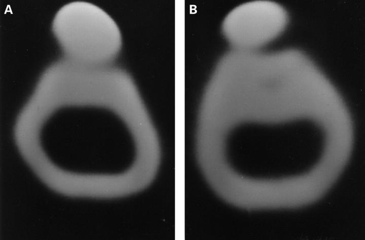
Figure 1 .
Postoperative CT findings. (A) Control. (B) Lateral shifting of patella and narrowing of lateral joint space in subluxation group.
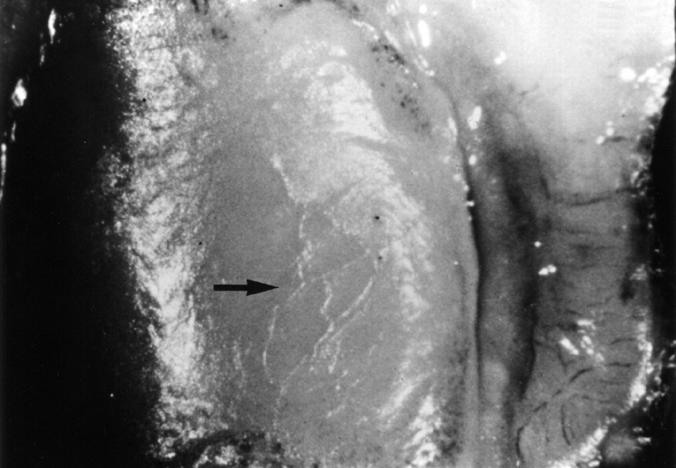
Figure 2 .
Macroscopic findings in patellar cartilage at eight weeks. Softening, swelling, and irregularity (arrows) are noted on surface of patellar cartilage.
Figure 3 .
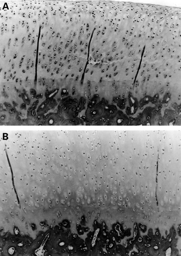
(A) Histological changes in patellar cartilage at eight weeks. Horizontal splits and oedematous changes in matrix in intermediate zones were evident. Surface of cartilage is smooth and no changes in chondrocytes were noted in superficial zone. (Safranin-O, original magnification × 100). (B) Patellar articular cartilage of control patellae at eight weeks. No remarkable change was noted. (Safranin-O, original magnification × 120).
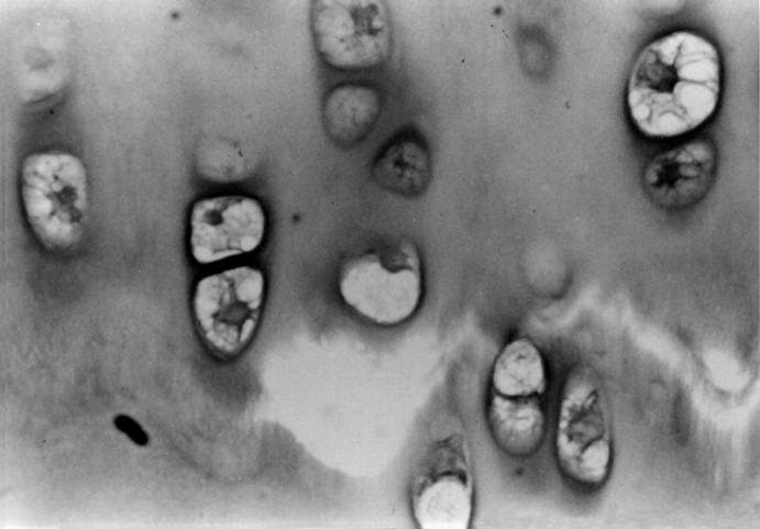
Figure 4 .
Histological changes around horizontal splits in intermediate zone at eight weeks. In vicinity of splits, chondrocytes of various sizes and with a number of vacuoles in cytoplasm were evident as well as reduction in matrix Safranin-O staining. (Safranin-O, original magnification × 200).
Figure 5 .
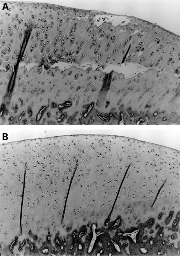
(A) At 12 weeks, horizontal splits have expanded widely and transversely parallel to surface with blister formation on articular surface and reduction in Safranin-O staining around blister is evident. Morphology of chondrocytes in superficial layer and deep zone differs. (Safranin-O, original magnification × 100). (B) Patellar articular cartilage of control side at 12 weeks. No remarkable change is noted in chondrocytes matrix. (Safranin-O, original magnification × 100).
Figure 6 .
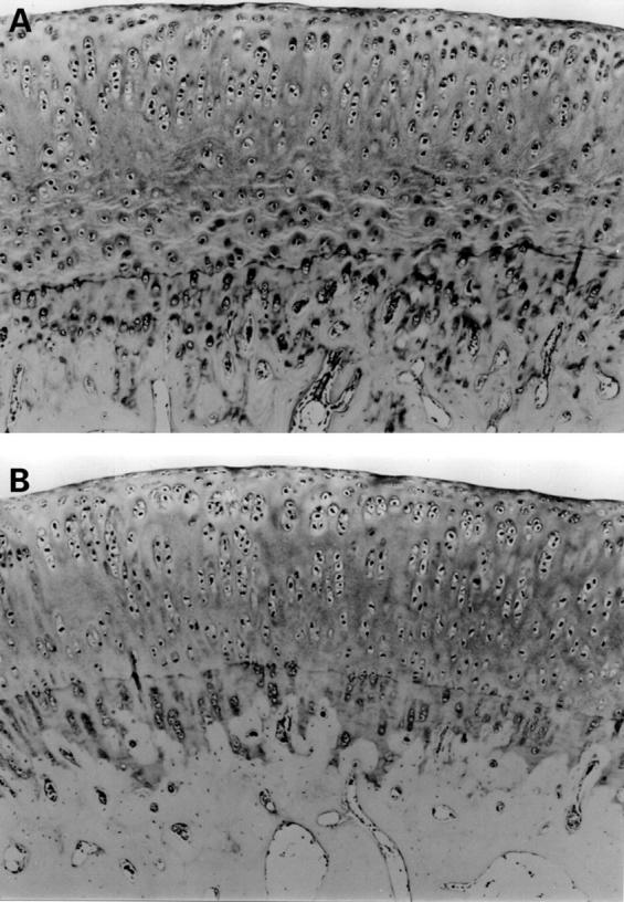
(A) At 32 weeks, cartilage is oedematous and many splits and wrinkles of matrix are evident in intermediate zones of patellar. Chondrocytes of superficial zone are not morphologically different to chondrocytes of control. (Safranin-O, original magnification × 100). (B) Patellar cartilage at 32 weeks in control. No remarkable changes noted in chondrocytes and matrix. (Safranin-O, original magnification × 100).
Selected References
These references are in PubMed. This may not be the complete list of references from this article.
- Dandy D. J., Poirier H. Chondromalacia and the unstable patella. Acta Orthop Scand. 1975 Sep;46(4):695–699. doi: 10.3109/17453677508989252. [DOI] [PubMed] [Google Scholar]
- Darracott J., Vernon-Roberts B. The bony changes in "chondromalacia patellae". Rheumatol Phys Med. 1971 Nov;11(4):175–179. doi: 10.1093/rheumatology/11.4.175. [DOI] [PubMed] [Google Scholar]
- Goodfellow J., Hungerford D. S., Woods C. Patello-femoral joint mechanics and pathology. 2. Chondromalacia patellae. J Bone Joint Surg Br. 1976 Aug;58(3):291–299. doi: 10.1302/0301-620X.58B3.956244. [DOI] [PubMed] [Google Scholar]
- Imai N., Tomatsu T., Okamoto H., Nakamura Y. Clinical and roentgenological studies on malalignment disorders of the patello-femoral joint. Part III: Lesions of the patellar cartilage and subchondral bone associated with patello-femoral malalignment. Nihon Seikeigeka Gakkai Zasshi. 1989 Jan;63(1):1–17. [PubMed] [Google Scholar]
- LaBerge M., Audet J., Drouin G., Rivard C. H. Structural and in vivo mechanical characterization of canine patellar cartilage: a closed chondromalacia patellae model. J Invest Surg. 1993 Mar-Apr;6(2):105–116. doi: 10.3109/08941939309141602. [DOI] [PubMed] [Google Scholar]
- Mankin H. J., Dorfman H., Lippiello L., Zarins A. Biochemical and metabolic abnormalities in articular cartilage from osteo-arthritic human hips. II. Correlation of morphology with biochemical and metabolic data. J Bone Joint Surg Am. 1971 Apr;53(3):523–537. [PubMed] [Google Scholar]
- Meachim G., Bentley G. Horizontal splitting in patellar articular cartilage. Arthritis Rheum. 1978 Jul-Aug;21(6):669–674. doi: 10.1002/art.1780210610. [DOI] [PubMed] [Google Scholar]
- Møller B. N., Møller-Larsen F., Frich L. H. Chondromalacia induced by patellar subluxation in the rabbit. Acta Orthop Scand. 1989 Apr;60(2):188–191. doi: 10.3109/17453678909149251. [DOI] [PubMed] [Google Scholar]
- Ohno O., Naito J., Iguchi T., Ishikawa H., Hirohata K., Cooke T. D. An electron microscopic study of early pathology in chondromalacia of the patella. J Bone Joint Surg Am. 1988 Jul;70(6):883–899. [PubMed] [Google Scholar]
- Saito S., Ryu J., Yamamoto K., Kohno H. [An experimental study on early changes of articular cartilages in subluxated patella of rabbits]. Nihon Seikeigeka Gakkai Zasshi. 1991 Aug;65(8):571–579. [PubMed] [Google Scholar]
- Thompson R. C., Jr, Oegema T. R., Jr, Lewis J. L., Wallace L. Osteoarthrotic changes after acute transarticular load. An animal model. J Bone Joint Surg Am. 1991 Aug;73(7):990–1001. [PubMed] [Google Scholar]
- WILES P., ANDREWS P. S., DEVAS M. B. Chondromalacia of the patella. J Bone Joint Surg Br. 1956 Feb;38-B(1):95–113. doi: 10.1302/0301-620X.38B1.95. [DOI] [PubMed] [Google Scholar]


