Abstract
OBJECTIVE—To report on four patients with Behçet's disease associated with endomyocardial fibrosis involving the right or the left ventricle. METHODS—Charts of more than 350 patients with Behçet's disease were reviewed. Endomyocardial fibrosis was confirmed because of cardiac failure in three patients and incidentally discovered by histological examination of an operative specimen in one patient. Echocardiography displayed bright echogen endocardium. Angiocardiography showed a reduced ventricular size. Electron beam computed tomography demonstrated a lowdense area involving the endocardium. Magnetic resonance imaging showed a mass of intermediate intensity on T1 weighted images. Diagnosis of endomyocardial fibrosis was based on histological study of a biopsy specimen in one patient and of an operative specimen in three. RESULTS—Six other similar cases of endomyocardial fibrosis complicating Behçet's disease were previously reported in the medical literature. Endomyocardial fibrosis predominantly involved the right ventricle. It can be considered a feature of Behçet's disease because: (a) no other cause was discovered; (b) arteritis, valvulopathy, and intraventricular thrombus were closely linked, and (c) all patients with endomyocardial fibrosis had vasculo-Behçet pattern. CONCLUSION—Endomyocardial fibrosis may be the sequelae of vasculitis involving endocardium or myocardium, or both and complicated with intraventricular thrombosis. Behçet's disease should be added to the list of causes of endomyocardial fibrosis.
Full Text
The Full Text of this article is available as a PDF (112.2 KB).
Figure 1 .
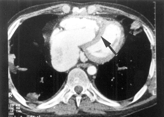
Electron beam computed tomography: low dense apical area involving the right ventricle.
Figure 2 .
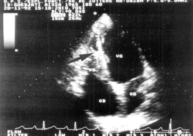
Four chamber bidimensional end diastolic echocardiogram: bright echoes in the right ventricular endocardium.
Figure 3 .

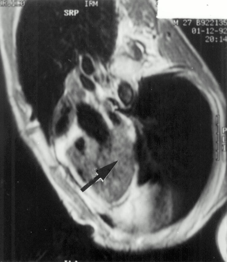
Magnetic resonance imaging: intermediate signal intensity mass on T1 weighted image occupying the middle part of the right ventricle.
Figure 4 .
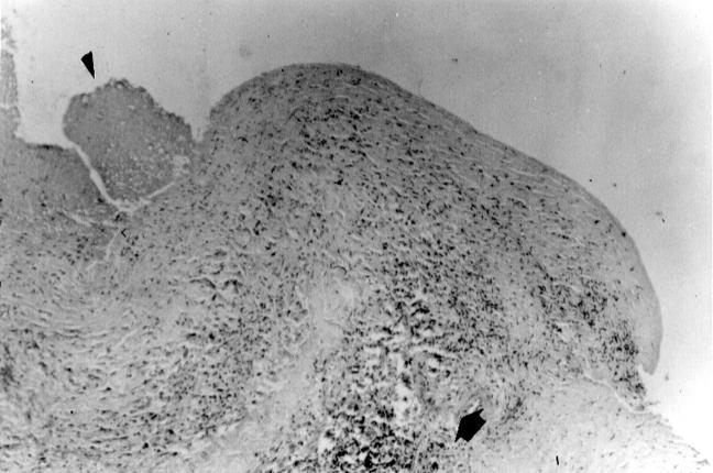
Histological examination of the operative endomyocardial fibrosis specimen (Haematein eosin safran, original magnification × 5): dense fibrous tissue with neovessels, granulocytes, mononuclear cells and fibroblasts infiltrate and anchored thrombus.


