Abstract
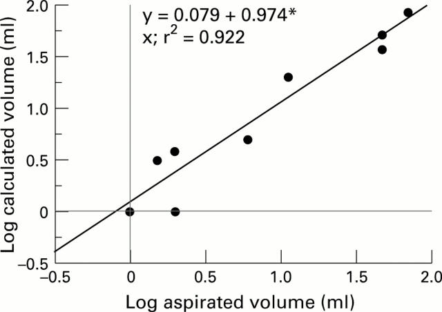
OBJECTIVES—To investigate the potential of quantitative magnetic resonance imaging (MRI) to differentiate between therapeutically induced changes in inflammation and synovial proliferation in rheumatoid arthritis (RA) of the knee. METHODS—MRI of the knee was performed on patients with RA before and one week after injection with corticosteroid (triamcinolone acetonide, TA group, n=9) and before, four, and 12 weeks after injection with yttrium-90 plus TA (TA+Y group, n=7). MRI scans were analysed by subjective visual grading by a trained observer and by computer aided quantitation for three features: synovial fluid volume, synovial pannus volume, and synovial enhancement after intravenous contrast agent. RESULTS—All TA subjects improved clinically at one week but the effects of TA+Y were more variable. TA significantly reduced synovial enhancement and effusion volume, whereas TA+Y at 12 weeks tended to increase synovial enhancement and decrease pannus volume. Quantitative MRI values agreed well with subjective assessment of scans. Comparison of calculated change on MRI scan before and immediately after aspiration with actual volume aspirated showed high correlation (r=0.96). CONCLUSIONS—Quantitative MRI correlates with subjective visual assessment and, at least for synovial fluid, is accurate. MRI can differentiate actions of two therapeutic modalities on various pathological processes and is sensitive enough to detect change after one week. With the additional advantage of lack of observer bias, it will probably become a useful tool in the development and assessment of existing and novel treatments.
Full Text
The Full Text of this article is available as a PDF (99.3 KB).
Figure 1 .
Comparison of aspirated volume of synovial fluid with calculated change in volume on MRI. Log transformations used. r = 0.9604.


